. 2017 Dec 6;25(12):2743-2752.
doi: 10.1016/j.ymthe.2017.08.007. Epub 2017 Aug 12.

Guanidinylated Neomycin Conjugation Enhances Intranasal Enzyme Replacement in the Brain

Affiliations

Guanidinylated Neomycin Conjugation Enhances Intranasal Enzyme Replacement in the Brain

Wenyong Tong et al. Mol Ther. .

Abstract

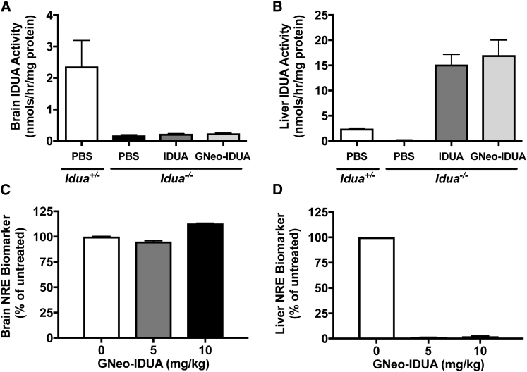
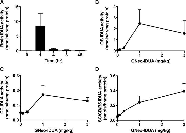
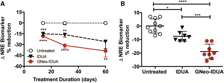
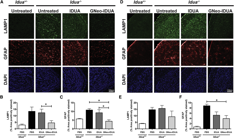
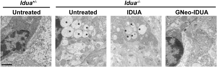
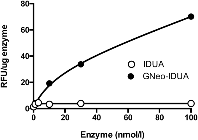
Iduronidase (IDUA)-deficient mice accumulate glycosaminoglycans in cells and tissues and exhibit many of the same neuropathological symptoms of patients suffering from Mucopolysaccharidosis I. Intravenous enzyme-replacement therapy for Mucopolysaccharidosis I ameliorates glycosaminoglycan storage and many of the somatic aspects of the disease but fails to treat neurological symptoms due to poor transport across the blood-brain barrier. In this study, we examined the delivery of IDUA conjugated to guanidinoneomycin (GNeo), a molecular transporter. GNeo-IDUA and IDUA injected intravenously resulted in reduced hepatic glycosaminoglycan accumulation but had no effect in the brain due to fast clearance from the circulation. In contrast, intranasally administered GNeo-IDUA entered the brain rapidly. Repetitive intranasal treatment with GNeo-IDUA reduced glycosaminoglycan storage, lysosome size and number, and neurodegenerative astrogliosis in the olfactory bulb and primary somatosensory cortex, whereas IDUA was less effective. The enhanced efficacy of GNeo-IDUA was not the result of increased nose-to-brain delivery or enzyme stability, but rather due to more efficient uptake into neurons and astrocytes. GNeo conjugation also enhanced glycosaminoglycan clearance by intranasally delivered sulfamidase to the brain of sulfamidase-deficient mice, a model of Mucopolysaccharidosis IIIA. These findings suggest the general utility of the guanidinoglycoside-based delivery system for restoring missing lysosomal enzymes in the brain.

Keywords: enzyme-replacement therapy; guanidinoglycosides; intranasal delivery; mucopolysaccharidoses; neuropathology.


Figures

Figure 1
Figure 1
Intravenous Administration of GNeo-Conjugated IDUA Shows Delivery Restricted to Somatic Tissues MPSI mice (Idua/) were treated intravenously with 5 mg/kg GNeo-IDUA, unconjugated IDUA, or PBS. Tissues were harvested 1 hr following treatment. (A) IDUA enzyme activity in whole brains. (B) IDUA enzyme activity in livers. (C) Non-reducing end heparan sulfate analysis by liquid chromatography/mass spectrometry in whole brains 48 hr after injection of enzyme. (D) Non-reducing end heparan sulfate analysis in livers. n = 2–3 mice per bar. Error bars indicate SEM.
Figure 2
Figure 2
Pharmacokinetic and Biodistribution Analysis of Intranasally Administered GNeo-IDUA in the Brain (A) Idua/ mice were treated intranasally with 1 mg/kg GNeo-IDUA. IDUA enzyme activity was measured at 0, 1, 4, 8, and 48 hr following treatment. n = 2–4 mice per bar. (B–D) Idua/ mice were treated intranasally with the indicated concentrations of GNeo-IDUA, and different brain regions were collected 1 hr following treatment. n = 3 mice per data point. (B) IDUA enzyme activity in the olfactory bulbs. (C) IDUA enzyme activity in the cerebral cortex (CC). (D) IDUA enzyme activity in subcortical (SC), cerebellum (CB), and brain stem (BS) regions. Error bars indicate SEM.
Figure 3
Figure 3
GNeo-IDUA Reduces the NRE Heparan Sulfate Biomarker in the MPSI Mouse Brain (A) Idua/ mice were treated intranasally with 1 mg/kg IDUA or GNeo-IDUA every other day for 14, 30, and 60 days. n = 10–12 mice for 14 days and 30 days; n = 5–6 mice for 60 days. (B) Idua/ mice were treated intranasally with 1 mg/kg IDUA or GNeo-IDUA twice a day for 14 days. n = 7–10 mice per treatment group from two separate experiments. Non-reducing end biomarker was determined in heparan sulfate isolated from whole brains. *p < 0.05, **p < 0.01, ***p < 0.001, ****p < 0.0001. Error bars indicate SEM, and significance was determined by one-way analysis of variance (ANOVA) with Tukey’s post-hoc analysis.
Figure 4
Figure 4
GNeo-IDUA Reduces Histopathological Hallmarks in the Aged MPSI Mouse Brain Idua/ mice were treated intranasally with 1 mg/kg IDUA or GNeo-IDUA twice a day for 30 days and then once a day for 30 days. Coronal sections were taken from different brain regions and stained with antibodies against LAMP1, a lysosomal marker, or GFAP, a marker of astrocytosis. (A) Representative confocal images acquired from olfactory bulbs. (B) Quantification of LAMP1 and (C) GFAP in the granule cell layer of the olfactory bulb. (D) Representative confocal images acquired from the primary somatosensory cortex. (E) Quantification of LAMP1 and (F) GFAP in layer II/III of the primary somatosensory cortex. n = 3 animals per treatment group. Error bars indicate SEM, and significance was determined by one-way analysis of variance (ANOVA) with Tukey’s post-hoc analysis.
Figure 5
Figure 5
GNeo-IDUA Reduces Lysosomal Storage in the MPSI Cerebral Cortex Electron microscopy images show that the number of lysosomal storage vesicles (asterisks) is reduced following GNeo-IDUA treatment. Scale bar, 1 μm.
Figure 6
Figure 6
Enhanced Uptake of GNeo-IDUA in Rat Neurons Cultures of primary rat cortical neurons and astrocytes were treated with the indicated concentration of IDUA or GNeo-IDUA for 2 hr. The cells were washed, trypsin treated, sedimented by centrifugation, washed twice, and subsequently assayed for iduronidase activity. Each data point represents a single well. The experiment was performed twice with comparable findings.

Similar articles

Cited by

References

    1. Esko J.D., Selleck S.B. Order out of chaos: assembly of ligand binding sites in heparan sulfate. Annu. Rev. Biochem. 2002;71:435–471. - PubMed
    1. Neufeld E.F., Muenzer J. The mucopolysaccharidoses. In: Scriver C.R., Sly W.S., Childs B., Beaudet A.L., Valle D., Kinzler K.W., Vogelstein B., editors. Eighth Edition. Volume 3. MacGraw-Hill; 2001. pp. 3421–3452. (Metabolic and Molecular Basis of Inherited Disease).
    1. Dwyer C.A., Scudder S.L., Lin Y., Dozier L.E., Phan D., Allen N.J., Patrick G.N., Esko J.D. Neurodevelopmental changes in excitatory synaptic structure and function in the cerebral cortex of Sanfilippo syndrome IIIA mice. Sci. Rep. 2017;7:46576. - PMC - PubMed
    1. Kelly J.M., Bradbury A., Martin D.R., Byrne M.E. Emerging therapies for neuropathic lysosomal storage disorders. Prog. Neurobiol. 2017;152:166–180. - PubMed
    1. Oh D.B. Glyco-engineering strategies for the development of therapeutic enzymes with improved efficacy for the treatment of lysosomal storage diseases. BMB Rep. 2015;48:438–444. - PMC - PubMed

MeSH terms