. 2018 Jan 1:368:171-186.
doi: 10.1016/j.neuroscience.2017.09.031. Epub 2017 Sep 27.

3D reconstruction and standardization of the rat facial nucleus for precise mapping of vibrissal motor networks

Affiliations

3D reconstruction and standardization of the rat facial nucleus for precise mapping of vibrissal motor networks

Jason M Guest et al. Neuroscience. .

Abstract

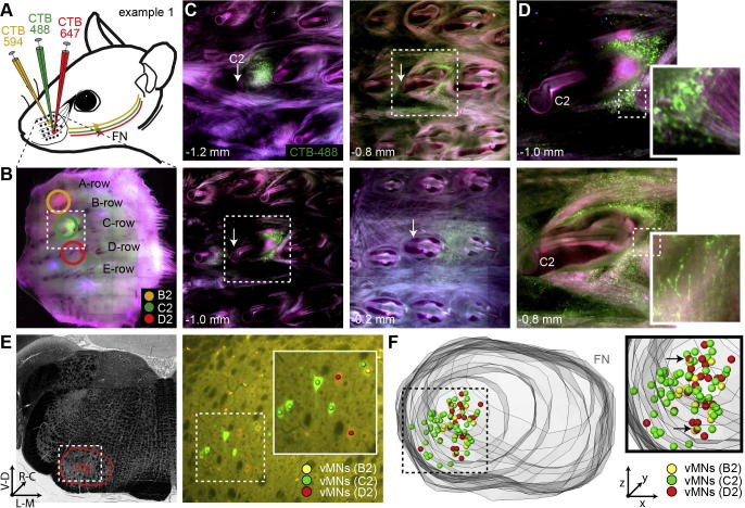
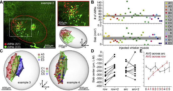
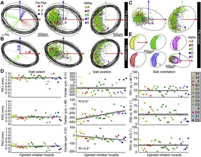
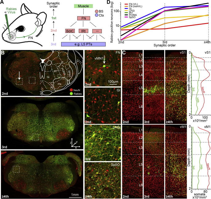
The rodent facial nucleus (FN) comprises motoneurons (MNs) that control the facial musculature. In the lateral part of the FN, populations of vibrissal motoneurons (vMNs) innervate two groups of muscles that generate movements of the whiskers. Vibrissal MNs thus represent the terminal point of the neuronal networks that generate rhythmic whisking during exploratory behaviors and that modify whisker movements based on sensory-motor feedback during tactile-based perception. Here, we combined retrograde tracer injections into whisker-specific muscles, with large-scale immunohistochemistry and digital reconstructions to generate an average model of the rat FN. The model incorporates measurements of the FN geometry, its cellular organization and a whisker row-specific map formed by vMNs. Furthermore, the model provides a digital 3D reference frame that allows registering structural data - obtained across scales and animals - into a common coordinate system with a precision of ∼60 µm. We illustrate the registration method by injecting replication competent rabies virus into the muscle of a single whisker. Retrograde transport of the virus to vMNs enabled reconstruction of their dendrites. Subsequent trans-synaptic transport enabled mapping the presynaptic neurons of the reconstructed vMNs. Registration of these data to the FN reference frame provides a first account of the morphological and synaptic input variability within a population of vMNs that innervate the same muscle.

Keywords: brain stem; connectivity; rabies virus; whisker; whisking.


Figures

Fig. 1
Fig. 1
Geometry and cellular organization of rat FN. (A) Schematic illustration of the musculature underlying whisker motor control (adapted from (Berg and Kleinfeld, 2003)). Injections of retrograde (CTB) and trans-synaptic (rabies virus) tracers into whisker-specific intrinsic muscles were performed to reveal the organization vMNs in rat FN. (B) Maximum projection image across all histological brain sections that comprise the FN. Sections were labeled with NeuN to reveal neuron somata. (C) Zoom-into one optical section from the image stack in panel (B). The outlines of the FN were identified by differences in soma size. Bottom panels show zoom-into illustrate the criteria to delineate the FN by NeuN-labeling. The center locations of all somata within the FN were marked (green spheres). (D) Result of reconstructing the FN outlines and marking all somata from the example experiment shown in panels (B, C). (E) The outlines were transformed into a surface that delineates the volume of the FN. The soma counts were transformed into a 3D density at 50-µm3 resolution. (F) The shape and volume of the FN were approximated by an ellipsoid. The experiment shown in panels (B–E) was repeated for two additional rats and the three FN reconstructions were aligned to yield an average ellipsoid (blue), which represents the FN reference frame. (G) Average (±SDs) volume and soma density of the FN model.
Fig. 2
Fig. 2
Labeling of whisker-specific vMNs in rat FN. (A) Schematic illustration of the injection experiments. Injections of three different versions of a retrograde tracer (i.e. CTB-488, 594 and 647) were targeted to the intrinsic muscles of adjacent whiskers (N = 6). (B–F) Illustrations of the injection sites and quantifications of the retrogradely labeled vMNs for one exemplary experiment, in which injections were targeted to the B2, C2 and D2 whiskers, respectively. (B) Example image of the mystacial pad showing the injections sites. (C) The pad shown in panel (B) was sliced into consecutive 150-µm-thick sections. Top-left panel (1.2 mm underneath the surface of the pad) shows the center location of the injection, which is posterior to the base of the follicle (white arrow) of the targeted C2 whisker. The remaining three panels illustrate that injections did not spread into the follicle and adjacent intrinsic (see panels at 0.8 and 1.0 mm depths) or extrinsic (see panel at 0.2 mm depth) muscles. (D) Zoom-ins to panels in (C) illustrate that the retrograde tracer spreads within the injected muscle, i.e. the shape of the injection site resembles the sling of the injected intrinsic muscle. (E) Left panel: Bright field image of 50-µm-thick coronal section allows delineating the outlines of the FN. Right panel: Widefield fluorescent images (superimposed with the bright field image) illustrate that each CTB injection (i.e. corresponding to panels (B–D)) labels a specific set of vMNs. (F) 3D reconstruction of the FN and distributions of all retrogradely labeled vMNs. The zoom-in shows that vMNs were largely whisker-specific and confined to the ventrolateral part of the FN. Note: The black arrows denote the vMNs that were labeled by two retrograde markers (i.e. C2/D2), likely reflecting the small spread of the C2 injection toward the D2 whisker (see panel (C)).
Fig. 3
Fig. 3
Organization of vMNs in rat FN. (A) Example of one of the 13 experiments in which we injected CTB into intrinsic muscles that were separated by (at least) one whisker. Here, injections were targeted to the intrinsic muscle of the A1 and C3 whiskers, respectively. Left panel: Maximum projection confocal image across all histological brain sections that comprised the FN. Vibrissal MNs for both injected whiskers were found throughout the FN along the R–C axis. Vibrissal MNs were whisker-specific and confined to the ventrolateral part. Right panels show zoom-in (top) and the center locations of all retrogradely labeled vMNs (bottom). The volumes occupied by the respective vMN distributions (i.e. shell-shaped slabs) were approximated by calculating a convex hull around the marked somata (light red and green surfaces). (B) The numbers of retrogradely labeled vMNs (top) and the respective slab volumes for all injected whisker muscles (n = 36). The black lines and gray-shaded areas represent mean and SEM, respectively. (C) Reconstruction of retrogradely labeled vMNs and slab volumes for two additional example experiments. Here, injections were targeted to the intrinsic muscles of the A3, C3 and E3 whisker (left panel), and to the C1, C3 and C5 whisker (right panel). Vibrissal MNs representing whiskers within the same row are disjoint, but located within the same slab. (D) Quantification of the slab positions along the L–M axis within each individual experiment (n = 36 injections in N = 13 rats). Slabs representing different whisker rows were always shifted with respect to each other. Slabs representing different whisker arcs (i.e. within the same row) showed no systematic shift with respect to each other (two-sided T-test: p < 0.01). (E) Quantification of the average slab locations along the L–M axis. Error bars denote mean ± SEM.
Fig. 4
Fig. 4
Standardized FN reference frame with vMN slabs. (A–C). The FN of all CTB-injected animals (N = 13) were approximated by an ellipsoid, whose centers of symmetry were aligned to the one of the FN reference frame (panel (A)), whose PAs were rotated to match the respective ones of the FN reference frame (panel (B)) and whose dimensions were linearly scaled to match the respective dimensions of the FN reference frame (panel (C)). In all panels, the respective distributions of retrogradely labeled somata were transformed accordingly. (D) After registration, each vMN slab was approximated by an ellipsoid (n = 36) and their respective geometrical parameters are plotted after sorting them by whisker row and then by arc (i.e. from left to right: A1–E3). The black lines and gray-shaded areas represent the mean and SEM of the respective parameter in each panel. Left: The 3D extent of the ellipsoids (i.e. length of the three PAs) did not correlate with the location of a particular whisker along the row or arc. Center: The slab ellipsoids’ center locations along the L–M and V–D axes (but not along the R–C axis) did correlate with the location of a particular whisker along the row (but not along the arc). Right: The 3D orientations of the slab ellipsoids did also not correlate with the location of a particular whisker along the row or arc. (E) Geometrical reference frame of the rat FN including an average map of whisker row-specific slabs. Slab positions represent the mean of the respective parameters shown in panel (D). Slab sizes and orientations are equal for all slabs and based on the C-row slab (see Experimental Procedures).
Fig. 5
Fig. 5
Trans-synaptic tracing of whisker muscle-related neuronal networks. (A) Schematic illustration of injection experiments. The N2c strain of rabies virus was injected into the C3 whisker muscle. Right panel: Illustration of defining the trans-synaptic order of rabies labeling. The virus labels vMNs in the FN, then neurons that are presynaptic to those first-order neurons at the level of the brain stem (BS, 2nd order neurons). Third order experiments can be identified by additional labeling that is specific to L5 of the cortex. Forth order experiments show cortical labeling in additional layers. (B) Example images (from top to bottom) represent 2nd (no labeling in cortex), third- (cortical labeling is restricted to L5) and ≥fourth-(cortical labeling in all layers) order spread (see panel (C)). Right panels: within each experiment, a sparse population of vMNs is labeled within the FN, whereas labeling increases with order in different BS nuclei such as the Gi, LPGi and Sp5O (see Table 1 for a list of abbreviations). (C) Example images (left panels) and density distributions of rabies-positive neurons in cortex (right panels) for experiments with second-, third-and ≥fourth-order trans-synaptic spread (images correspond to the brain stem images in panel (B)). Each of the four density profiles (green) represents average from manual quantifications of rabies-positive neurons in three coronal sections through vS1 and vM1, respectively. (D) Quantification of all rabies-positive neurons within the BS depending on the order of rabies spread. In all infected BS nuclei, the number of labeled neurons increases by one order of magnitude from 2nd to 3rd order. The increase from 3rd to ≥4th order is 2–fourfold. The number of rabies-positive neurons within the ventrolateral (VL) FN is independent of the order of spread, but increases outside the ventrolateral FN (nonVL).
Fig. 6
Fig. 6
Dendrite morphologies of vMNs. (A) Left panel: High-resolution image of the vMN shown in Fig.5B (i.e. vMN1; white arrow). The morphology of vMN1 was reconstructed across consecutive histological sections. Right panel: High-resolution image of histological section adjacent to the one shown in the left panel shows soma, dendrites and axon of a second vMN (i.e. vMN2) and the axon of vMN1 (white arrows). The zoom-in reveals rabies virus particles within the labeled cells. (B) 3D reconstruction of three vMNs from the same animal, including the two vMNs shown in panel (A). Note: Axons (black) of the reconstructed vMNs have no collaterals within the FN. (C) Registration of the rabies-labeled vMNs to the FN reference frame allows quantification of morphological variability with respect to the whisker row-specific map. (D) Quantification of dendrites within the FN reference frame reveal large morphological variability within the population of C3 innervating vMNs. Upper panel: the dendritic fields of the three C3 vMNs overlap only partially with each other (see also colored arrows in panel (C)). Lower panel: dendrites of vMNs extend beyond the FN. Within the FN, dendrites innervate the slabs in a cell-specific manner.
Fig. 7
Fig. 7
Presynaptic populations of vMNs. (A) Maximum confocal projection image across all histological brain sections that comprise the Sp5O of the BS in a second-order experiment after injecting rabies virus into the C3 whisker (same experiment as shown in Fig.5B, C). Images were superimposed with the corresponding pages from the Paxinos rat brain atlas and all rabies-positive neurons were marked and assigned to the respective BS nuclei. Right panel: zoom-into the GI reveals sparse labeling of second- order neurons that are presynaptic to the first-order vMNs in the FN, e.g. the ones in Fig. 6. (B) Maximum confocal projection image across all histological brain sections that comprise the Sp5I of the BS in same experiment as shown in panel (A) Right panel: zoom-into the IRt. (C) Maximum confocal projection image across all histological brain sections that comprise the Sp5C of the BS in the same experiment as shown in panels (A, B). Right panel: zoom-into the MdD. (D) Same BS regions as shown in the zoom-ins in panel (A) for a second second-order experiment after injections into the C3 whisker muscle. Labeling in the second animal was in general sparser than in the first experiment. (E) Quantification of the number of neurons in all rabies-infected BS nuclei in the two second-order C3 injection experiments. The red marked BS nuclei represent those that are consistently labeled in both experiments. The major sources of common input to vMNs are highlighted in green.

References

    1. Ashwell K.W. The adult mouse facial nerve nucleus: morphology and musculotopic organization. J Anat. 1982;135:531–538. - PMC - PubMed
    1. Berg R.W., Kleinfeld D. Vibrissa movement elicited by rhythmic electrical microstimulation to motor cortex in the aroused rat mimics exploratory whisking. J Neurophysiol. 2003;90:2950–2963. - PubMed
    1. Brecht M. What makes whiskers shake? Focus on Current flow in vibrissa motor cortex can phase-lock with exploratory rhythmic whisking in rat. J Neurophysiol. 2004;92:1265–1266. - PubMed
    1. Brecht M., Schneider M., Sakmann B., Margrie T.W. Whisker movements evoked by stimulation of single pyramidal cells in rat motor cortex. Nature. 2004;427:704–710. - PubMed
    1. Carvell G.E., Simons D.J., Lichtenstein S.H., Bryant P. Electromyographic activity of mystacial pad musculature during whisking behavior in the rat. Somatosens Mot Res. 1991;8:159–164. - PubMed