

Exosomes Derived from Dendritic Cells Treated with Schistosoma japonicum Soluble Egg Antigen Attenuate DSS-Induced Colitis
- PMID: 28959207
- PMCID: PMC5603738
- DOI: 10.3389/fphar.2017.00651
Exosomes Derived from Dendritic Cells Treated with Schistosoma japonicum Soluble Egg Antigen Attenuate DSS-Induced Colitis
Abstract
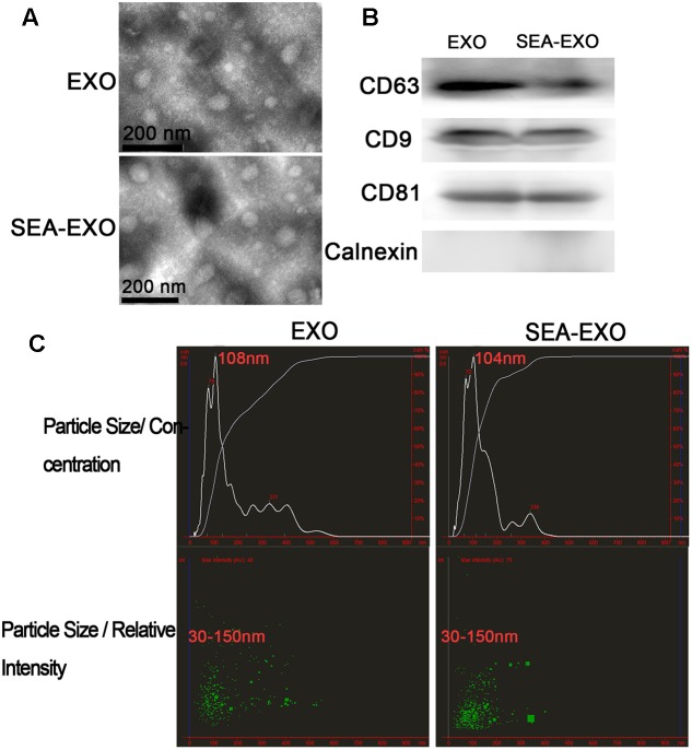
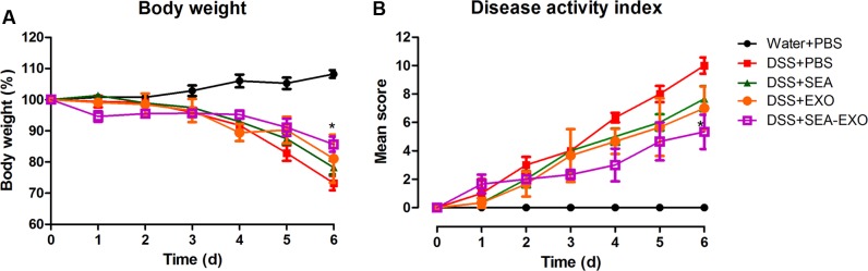
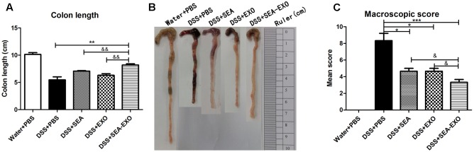
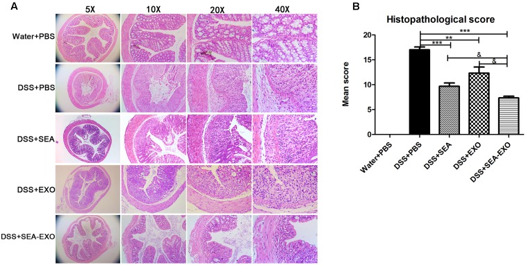
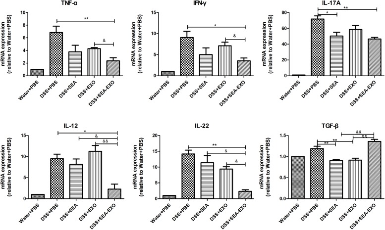
Exosomes are 30-150 nm small membrane vesicles that are released into the extracellular medium via cells that function as a mode of intercellular communication. Dendritic cell (DC)-derived exosomes modulate immune responses and prevent the development of autoimmune diseases. Moreover, Schistosoma japonicum eggs show modulatory effects in a mouse model of colitis. Therefore, we hypothesized that exosomes derived from DCs treated with S. japonicum soluble eggs antigen (SEA; SEA-treated DC exosomes) would be useful for treating inflammatory bowel disease (IBD). Exosomes were purified from the supernatant of DCs treated or untreated with SEA and identified via transmission electron microscopy, western blotting and NanoSight. Acute colitis was induced via the administration of dextran sulfate sodium (DSS) in drinking water (5.0%, wt/vol). Treatment with exosomes was conducted via intraperitoneal injection (i.p.; 50 μg per mouse) from day 0 to day 6. Clinical scores were calculated based on weight loss, stool type, and bleeding. Colon length was measured as an indirect marker of inflammation, and colon macroscopic characteristics were determined. Body weight loss and the disease activity index of DSS-induced colitis mice decreased significantly following treatment with SEA-treated DC exosomes. Moreover, the colon lengths of SEA-treated DC exosomes treated colitis mice improved, and their mean colon macroscopic scores decreased. In addition, histologic examinations and histological scores showed that SEA-treated DC exosomes prevented colon damage in acute DSS-induced colitis mice. These results indicate that SEA-treated DC exosomes attenuate the severity of acute DSS-induced colitis mice more effectively than DC exosomes. The current work suggests that SEA-treated DC exosomes may be useful as a new approach to treat IBD.
Keywords: dendritic cell; dextran sulfate sodium; exosomes; inflammatory bowel disease; soluble egg antigen.
Figures
Similar articles
-
Schistosoma japonicum attenuates dextran sodium sulfate-induced colitis in mice via reduction of endoplasmic reticulum stress.World J Gastroenterol. 2017 Aug 21;23(31):5700-5712. doi: 10.3748/wjg.v23.i31.5700. World J Gastroenterol. 2017. PMID: 28883695 Free PMC article.
-
Enterocyte dendritic cell-specific intercellular adhesion molecule-3-grabbing non-integrin expression in inflammatory bowel disease.World J Gastroenterol. 2015 Jan 7;21(1):187-95. doi: 10.3748/wjg.v21.i1.187. World J Gastroenterol. 2015. PMID: 25574091 Free PMC article.
-
Co-Stimulation-Impaired Bone Marrow-Derived Dendritic Cells Prevent Dextran Sodium Sulfate-Induced Colitis in Mice.Front Immunol. 2018 May 3;9:894. doi: 10.3389/fimmu.2018.00894. eCollection 2018. Front Immunol. 2018. PMID: 29774025 Free PMC article.
-
Exosomes Derived From M2b Macrophages Attenuate DSS-Induced Colitis.Front Immunol. 2019 Oct 16;10:2346. doi: 10.3389/fimmu.2019.02346. eCollection 2019. Front Immunol. 2019. PMID: 31749791 Free PMC article.
-
Regulatory T cell-derived exosomes: possible therapeutic and diagnostic tools in transplantation.Front Immunol. 2014 Nov 5;5:555. doi: 10.3389/fimmu.2014.00555. eCollection 2014. Front Immunol. 2014. PMID: 25414702 Free PMC article. Review.
Cited by
-
Schistosoma Infection and Schistosoma-Derived Products Modulate the Immune Responses Associated with Protection against Type 2 Diabetes.Front Immunol. 2018 Jan 17;8:1990. doi: 10.3389/fimmu.2017.01990. eCollection 2017. Front Immunol. 2018. PMID: 29387059 Free PMC article. Review.
-
Emerging role of exosomes in ulcerative colitis: Targeting NOD-like receptor family pyrin domain containing 3 inflammasome.World J Gastroenterol. 2024 Feb 14;30(6):527-541. doi: 10.3748/wjg.v30.i6.527. World J Gastroenterol. 2024. PMID: 38463022 Free PMC article. Review.
-
Herpes simplex virus interference with immunity: Focus on dendritic cells.Virulence. 2021 Dec;12(1):2583-2607. doi: 10.1080/21505594.2021.1980990. Virulence. 2021. PMID: 34895058 Free PMC article. Review.
-
Schistosoma japonicum Soluble Egg Antigen Protects Against Type 2 Diabetes in Lepr db/db Mice by Enhancing Regulatory T Cells and Th2 Cytokines.Front Immunol. 2019 Jun 26;10:1471. doi: 10.3389/fimmu.2019.01471. eCollection 2019. Front Immunol. 2019. PMID: 31297120 Free PMC article.
-
TNF-α and INF-γ primed canine stem cell-derived extracellular vesicles alleviate experimental murine colitis.Sci Rep. 2020 Feb 7;10(1):2115. doi: 10.1038/s41598-020-58909-4. Sci Rep. 2020. PMID: 32034203 Free PMC article.
References
-
- Boldeanu M. V., Silosi I., Ghilusi M., Cojocaru M., Biciusca V., Avramescu C. S., et al. (2014). Investigation of inflammatory activity in ulcerative colitis. Rom. J. Morphol. Embryol. 55 1345–1351. - PubMed
-
- Bonovas S., Fiorino G., Allocca M., Lytras T., Nikolopoulos G. K., Peyrin-Biroulet L., et al. (2016). Biologic therapies and risk of infection and malignancy in patients with inflammatory bowel disease: a systematic review and network meta-analysis. Clin. Gastroenterol. Hepatol. 14 1385–1397. 10.1016/j.cgh.2016.04.039 - DOI - PubMed
LinkOut - more resources
Full Text Sources
Other Literature Sources