

Role of lipocalin 2 in intraventricular haemoglobin-induced brain injury
- PMID: 28959462
- PMCID: PMC5435192
- DOI: 10.1136/svn-2016-000009
Role of lipocalin 2 in intraventricular haemoglobin-induced brain injury
Abstract
Objective: Our recent studies have shown that blood components, including haemoglobin and iron, contribute to hydrocephalus development and brain injury after intraventricular haemorrhage (IVH). The current study investigated the role of lipocalin 2 (LCN2), a protein involved in iron handling, in the ventricular dilation and neuroinflammation caused by brain injury in a mouse model of IVH.
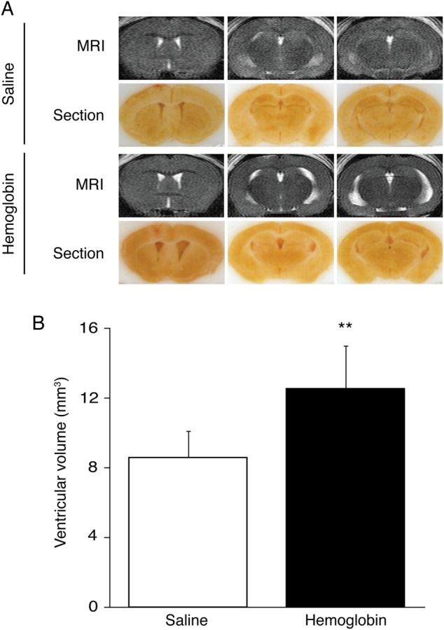
Design: Female wild-type (WT) C57BL/6 mice and LCN2-deficient (LCN2-/-) mice had an intraventricular injection of haemoglobin, and control mice received an equivalent amount of saline. MRI was performed presurgery and postsurgery to measure ventricular volume and the brains were used for either immunohistochemistry or western blot.
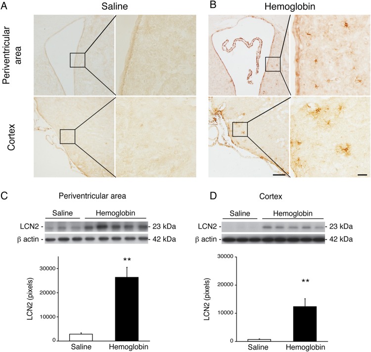
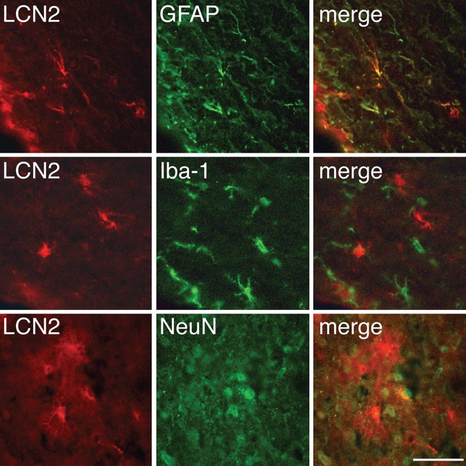
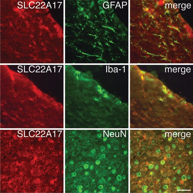
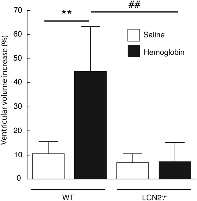
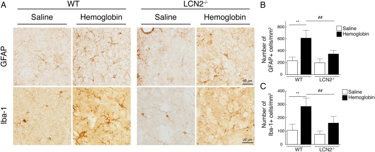
Results: Ventricular dilation was observed in WT mice at 24 h after haemoglobin (25 mg/mL, 20 µL) injection (12.5±2.4 vs 8.6±1.5 mm3 in the control, p<0.01). Western blotting showed that LCN2 was significantly upregulated in the periventricular area (p<0.01). LCN2 was mainly expressed in astrocytes, whereas the LCN2 receptor was detected in astrocytes, microglia/macrophages and neurons. Haemoglobin-induced ventricle dilation and glia activation were less in LCN2-/- mice (p<0.01). Injection of high-dose haemoglobin (50 mg/mL) resulted in lower mortality in LCN2-/- mice (27% vs 86% in WT; p<0.05).
Conclusions: Intraventricular haemoglobin caused LCN2 upregulation and ventricular dilation. Haemoglobin resulted in lower mortality and less ventricular dilation in LCN2-/- mice. These results suggest that LCN2 has a role in haemoglobin-induced brain injury and may be a therapeutic target for IVH.
Keywords: Hemorrhage; brain injury; hemoglobin; lipocalin 2; mouse.
Conflict of interest statement
Competing interests: None declared.
Figures
Similar articles
-
Role of lipocalin-2 in brain injury after intracerebral hemorrhage.J Cereb Blood Flow Metab. 2015 Sep;35(9):1454-61. doi: 10.1038/jcbfm.2015.52. Epub 2015 Apr 8. J Cereb Blood Flow Metab. 2015. PMID: 25853903 Free PMC article.
-
Lipocalin-2 Is a Key Regulator of Neuroinflammation in Secondary Traumatic and Ischemic Brain Injury.Neurotherapeutics. 2023 Apr;20(3):803-821. doi: 10.1007/s13311-022-01333-5. Epub 2022 Dec 12. Neurotherapeutics. 2023. PMID: 36508119 Free PMC article.
-
Lipocalin 2 and Blood-Brain Barrier Disruption in White Matter after Experimental Subarachnoid Hemorrhage.Acta Neurochir Suppl. 2016;121:131-4. doi: 10.1007/978-3-319-18497-5_23. Acta Neurochir Suppl. 2016. PMID: 26463936
-
Lipocalin-2 as a therapeutic target for brain injury: An astrocentric perspective.Prog Neurobiol. 2016 Sep;144:158-72. doi: 10.1016/j.pneurobio.2016.08.001. Epub 2016 Aug 4. Prog Neurobiol. 2016. PMID: 27498195 Review.
-
Role of lipocalin 2 in stroke.Neurobiol Dis. 2023 Apr;179:106044. doi: 10.1016/j.nbd.2023.106044. Epub 2023 Feb 17. Neurobiol Dis. 2023. PMID: 36804285 Review.
Cited by
-
Ablation of lipocalin-2 reduces neuroinflammation in a mouse model of Krabbe disease.Sci Rep. 2024 Dec 30;14(1):31822. doi: 10.1038/s41598-024-82927-1. Sci Rep. 2024. PMID: 39738378 Free PMC article.
-
Novel therapeutic modulators of astrocytes for hydrocephalus.Front Mol Neurosci. 2022 Sep 26;15:932955. doi: 10.3389/fnmol.2022.932955. eCollection 2022. Front Mol Neurosci. 2022. PMID: 36226316 Free PMC article. Review.
-
Complement Inhibition Reduces Early Erythrolysis, Attenuates Brain Injury, Hydrocephalus, and Iron Accumulation after Intraventricular Hemorrhage in Aged Rats.Transl Stroke Res. 2025 Jun;16(3):882-895. doi: 10.1007/s12975-024-01273-6. Epub 2024 Jun 28. Transl Stroke Res. 2025. PMID: 38943026
-
Posttraumatic hydrocephalus: Recent advances and new therapeutic strategies.Health Sci Rep. 2023 Nov 16;6(11):e1713. doi: 10.1002/hsr2.1713. eCollection 2023 Nov. Health Sci Rep. 2023. PMID: 38028696 Free PMC article.
-
CSF lipocalin-2 increases early in subarachnoid hemorrhage are associated with neuroinflammation and unfavorable outcome.J Cereb Blood Flow Metab. 2021 Oct;41(10):2524-2533. doi: 10.1177/0271678X211012110. Epub 2021 May 5. J Cereb Blood Flow Metab. 2021. PMID: 33951946 Free PMC article.
References
-
- Diringer MN, Edwards DF, Zazulia AR. Hydrocephalus: a previously unrecognized predictor of poor outcome from supratentorial intracerebral hemorrhage. Stroke 1998;29:1352–7. doi:10.1161/01.STR.29.7.1352 - DOI - PubMed
-
- Black PM. Hydrocephalus and vasospasm after subarachnoid hemorrhage from ruptured intracranial aneurysms. Neurosurgery 1986;18:12–16. doi:10.1227/00006123-198601000-00003 - DOI - PubMed
-
- Hasan D, Vermeulen M, Wijdicks EF, et al. . Management problems in acute hydrocephalus after subarachnoid hemorrhage. Stroke 1989;20:747–53. doi:10.1161/01.STR.20.6.747 - DOI - PubMed
-
- Dorai Z, Hynan LS, Kopitnik TA, et al. . Factors related to hydrocephalus after aneurysmal subarachnoid hemorrhage. Neurosurgery 2003;52:763–9; discussion 769-71 doi:10.1227/01.NEU.0000053222.74852.2D - DOI - PubMed
-
- Chen Z, Gao C, Hua Y, et al. . Role of iron in brain injury after intraventricular hemorrhage. Stroke 2011;42:465–70. doi:10.1161/STROKEAHA.110.602755 - DOI - PMC - PubMed
Publication types
MeSH terms
Substances
Grants and funding
LinkOut - more resources
Full Text Sources
Other Literature Sources
Miscellaneous