. 2015 Dec 17:3:78-86.
doi: 10.1016/j.toxrep.2015.12.004. eCollection 2016.

Diosmetin protects against retinal injury via reduction of DNA damage and oxidative stress

Affiliations

Diosmetin protects against retinal injury via reduction of DNA damage and oxidative stress

Zeren Shen et al. Toxicol Rep. .

Abstract

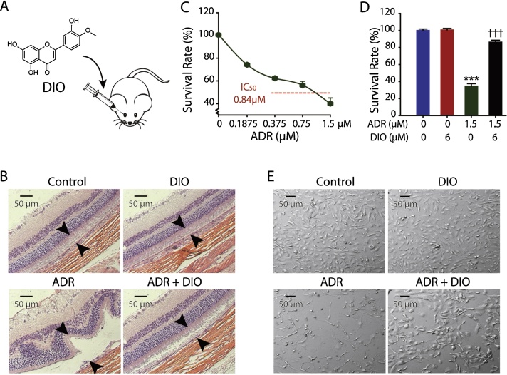
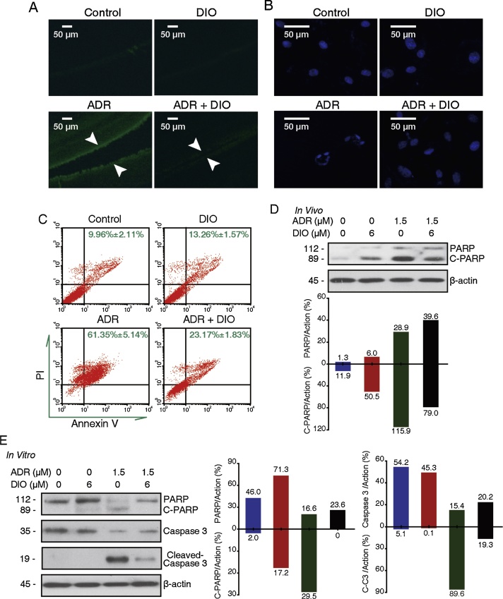
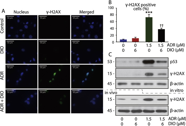
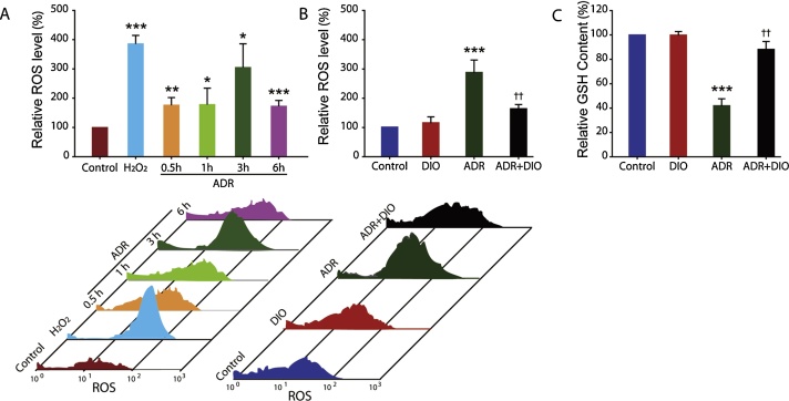
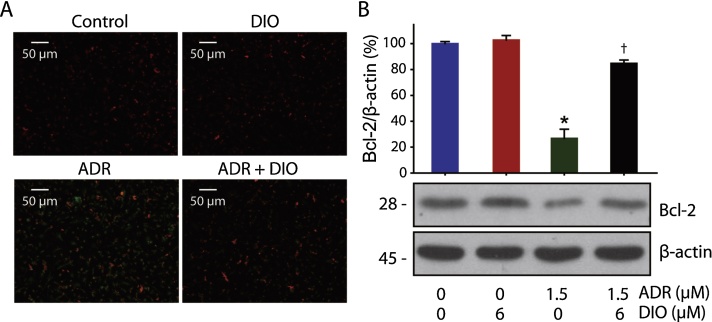
Visual impairment is a global public health problem that needs new candidate drugs. Chrysanthemum is a traditional Chinese drug, famous for its eye-protective function, with an unclear mechanism of action. To determine how chrysanthemum contributes to vision, we identified, for the first time, the component of chrysanthemum, diosmetin (DIO), which acts in protecting the injured retina in an adriamycin (ADR) improving model. We observed that DIO could attenuate the apoptosis of retinal cells in Sprague-Dawley rats and verified this effect in cultured human retinal pigment epithelium (RPE) cells, ARPE-19. Our further study on the mechanism revealed the counteractive effect of DIO on the attenuation of DNA damage and oxidative stress, which occurs in a wide range of retinal disorders. These results collectively promise the potential value of DIO as a retinal-protective agent for disorders that lead to blindness. In addition, we identified, for the first time, the component of chrysanthemum, DIO, which acts in protecting the injured retina.

Keywords: ADR, adriamycin; AMD, age-related macular degeneration; ATP, adenosine triphosphate; Apoptosis; CNV, choroidal neovascularisation; Chrysanthemum; DIO, diosmetin; DNA damage; Diosmetin; Diosmetin (PubChem CID5281612); Doxorubicin (PubChem CID31703); H&E, hematoxylin and eosin; IC50, inhibition for 50% of the cells; IVI, intravitreal injection; Oxidative stress; PVR, proliferative vitreoretinopathy; ROS, reactive oxygen species; RPE, retinal pigment epithelium; Retinal injury; Retinal pigment epithelium.


Figures

Fig. 1
Fig. 1
DIO protects against ADR-induced retinal injury in vivo and in vitro. (A) The chemical structure and the intravitreal injection (IVI) route of DIO. (B) Slices of retina were stained with hematoxylin and eosin (H&E) for histopathological analysis. Representative histomicrographs of retina sections of non-drug treament group, 6 μM DIO treatment group, 1.5 μM ADR treatment group and co-injected group. Arrows indicate the thickness of the pigment epithelium layer. (C) Determination of the IC50 of ARPE-19 cells treated with ADR at increasing concentrations (0–1.5 μM) for 72 h. The percentage of cell proliferation was measured with the MTT assay. (D) The cell survival rate was measured with MTT assays. Cells were treated with 6 μM of DIO and 1.5 μM of ADR for 72 h. The data represent the mean ± SD (n = 4), ***P < 0.001 (ADR vs. control), †††P < 0.001 (DIO + ADR vs. ADR). (E) Cell morphology was observed after 72 h of ADR (1.5 μM) and DIO (6 μM) treatment.
Fig. 2
Fig. 2
DIO attenuates the ADR-induced human retinal pigment epithelium cells apoptosis. (A) DIO reduced apoptosis in the RPE cells in vivo. The retinal tissues were terminal deoxynucleotidyl transferase-mediated dUTP nick end labelled and were imaged by fluorescent microscopy. The content of TUNEL-positive cells was equal to the number of green points in the photograph. Arrows indicate the thickness of the pigment epithelium layer. (B–C) DIO reduced the apoptosis of RPE cells in vitro. (B) ARPE-19 cells were treated with ADR and DIO for 72 h, and nuclei changes were photographed with fluorescence microscopy. (C) Flow cytometry recording shows the apoptosis rate of the ARPE-19 cells. (D) In vivo, retina extracts were analyzed by western blot analysis after IVI, PARP and cleaved PARP expression were analyzed. (E) In vitro, ARPE-19 cells were treated with drugs for 48 h and whole cell lysates were analyzed by western blot to evaluate the levels of caspase-3, PARP and their cleaved fragments.
Fig. 3
Fig. 3
DIO prevents ADR-triggered DNA damage in RPE cells. (A) Immunofluorescent micrographs of control and drug-treated (24 h) cells stained for γ-H2AX foci (blue—nucleus; green—γ-H2AX foci). (B) Quantitation of the γ-H2AX foci in the micrographs from A. The data represent the mean ± SD (n = 4), ***P < 0.001 (ADR vs. control), ††P < 0.01 (DIO + ADR vs. ADR). (C) In vitro western blot analysis of p53 and γ-H2AX expression in control and drug-treated (24 h) lysates. In vivo western blot analysis of γ-H2AX expression in control and drug-treated lysates.
Fig. 4
Fig. 4
DIO inhibits ADR-induced oxidative stress. (A) Flow cytometric evaluation of H2DCFDA stained-negative control, positive control and ADR-treated cells at various time points. (B) Effect of DIO on ADR (3 h) induced ROS in cultured RPE cells, as measured by flow cytometry. (C) The content of intracellular GSH after treatment with ADR and DIO for 12 h. The data represent the mean ± SD (n = 4), *P < 0.05, **P < 0.01, ***P < 0.001 (ADR vs. control), ††P < 0.01 (DIO + ADR vs. ADR).
Fig. 5
Fig. 5
DIO Inhibits ADR-induced mitochondria dysfunction in RPE cells. (A) The effect of DIO on loss of the mitochondrial membrane potential in RPE cells. After treatment with DIO and ADR for 24 h at the indicated concentrations, a loss in the mitochondrial membrane potential was observed by microscopy after JC-1 staining. The green fluorescence intensity indicated the cells with low mitochondrial membrane potential, while the red fluorescence intensity indicated the cells with stable mitochondrial membrane potential (n = 4). (B) Western blot analysis and corresponding densitometric measurements of the mitochondrial marker, Bcl-2. Effect of DIO on Bcl-2 protein expression induced by ADR in cultured RPE cells. Cells were incubated with 1.5 μM ADR and 6 μM DIO for 24 h and lysed; Bcl-2 was analyzed by western blot. The data represent the mean ± SD (n = 4), *P < 0.05 (ADR vs. control), P < 0.05 (DIO + ADR vs. ADR).

Similar articles

Cited by

References

    1. Abd-Alla H.I., Albalawy M.A., Aly H.F., Shalaby N.M.M., Shaker K.H. Flavone composition and antihypercholesterolemic and antihyperglycemic activities of Chrysanthemum coronarium L. Z. Naturforsch. C. 2014;69:199–208. - PubMed
    1. Banáth J.P., Olive P.L. Expression of phosphorylated histone H2AX as a surrogate of cell killing by drugs that create DNA double-strand breaks. Cancer Res. 2003;63:4347–4350. PubMed: 12907603. - PubMed
    1. Beatty S., Koh H.H., Phil M., Henson D., Boulton M. The role of oxidative stress in the pathogenesis of age-related macular degeneration. Surv. Ophthalmol. 2000;45:115–134. - PubMed
    1. Berlin V., Haseltine W.A. Reduction of adriamycin to a semiquinone-free radical by NADPH cytochrome P-450 reductase produces DNA cleavage in a reaction mediated by molecular oxygen. J. Biol. Chem. 1981;256:4747–4756. PubMed: 6262301. - PubMed
    1. Bordone M.P., Lanzani M.F., López-Costa J.J., Chianelli M.S., Franco P., Sáenz D.A., Rosenstein R.E. Bacterial lipopolysaccharide protects the retina from light-induced damage. J. Neurochem. 2012;122:392–403. - PubMed

LinkOut - more resources