

Toxicity determinants of multi-walled carbon nanotubes: The relationship between functionalization and agglomeration
- PMID: 28959543
- PMCID: PMC5615827
- DOI: 10.1016/j.toxrep.2016.01.011
Toxicity determinants of multi-walled carbon nanotubes: The relationship between functionalization and agglomeration
Abstract
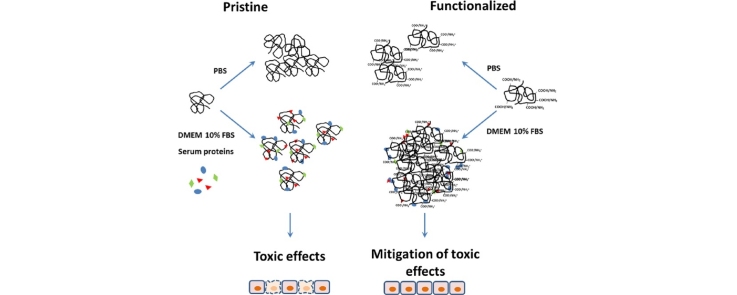
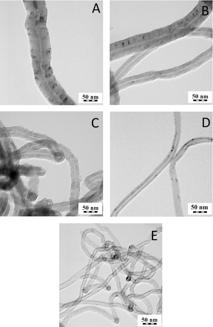
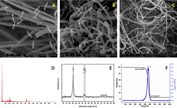
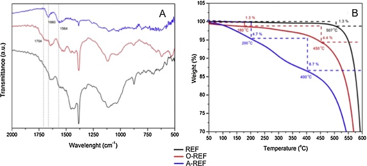
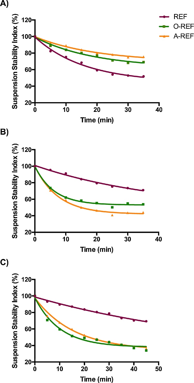
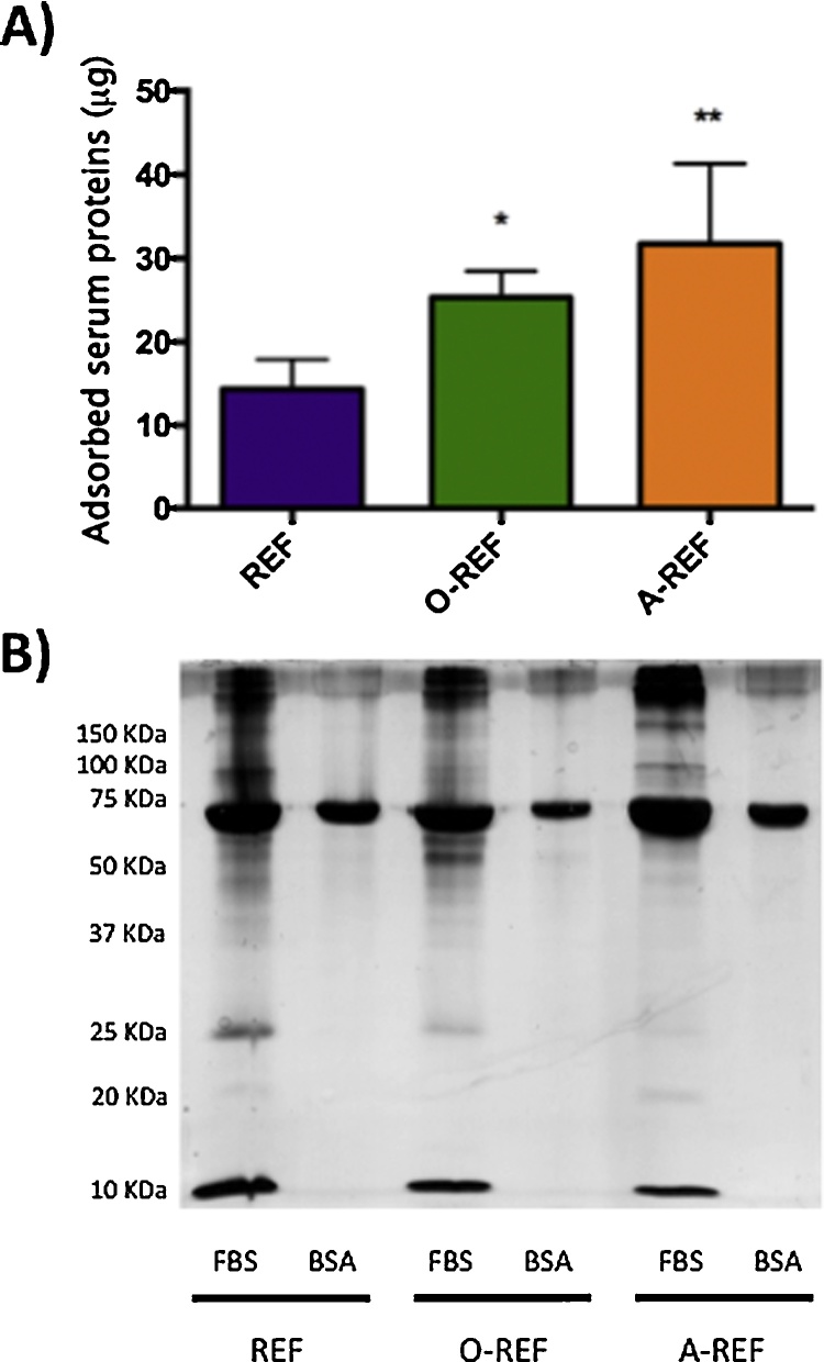
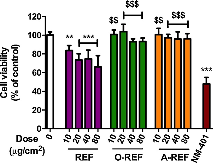
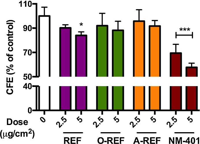
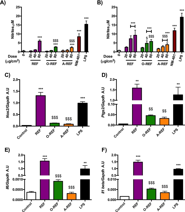
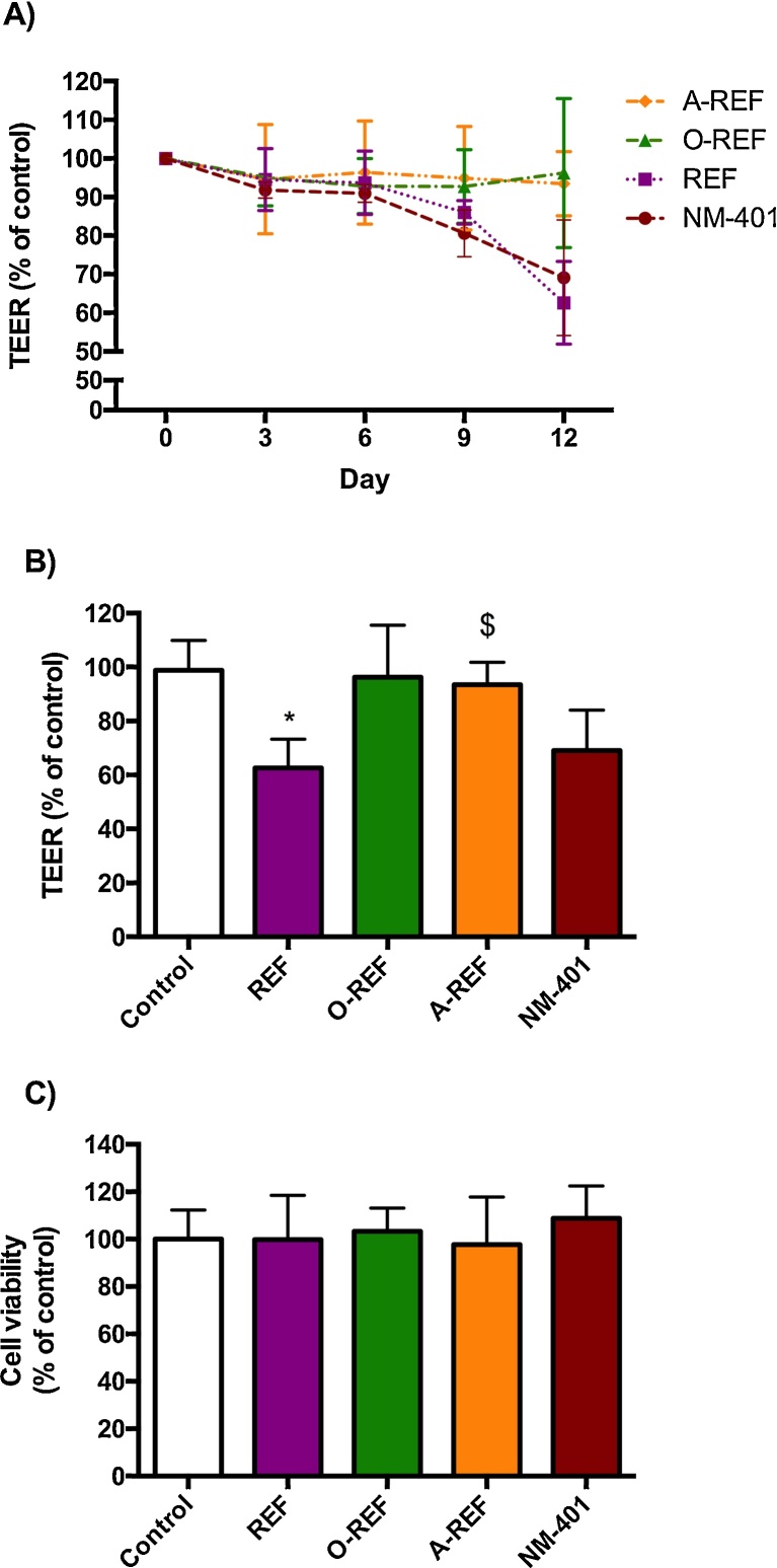
The elucidation of toxicity determinants of multi-walled carbon nanotubes (MWCNT) is still incomplete. Functionalization with carboxyl groups is, however, commonly used to mitigate MWCNT toxicity, although the rationale for the mitigating effect has not been fully clarified yet. In this work, two optimized chemical vapor deposition methods were employed to obtain MWCNT of comparable length but different diameter, which were subsequently functionalized. For MWCNT of diameter larger than 40 nm, no detrimental effects on cell viability of macrophages were observed, while mild cytotoxicity was recorded for diameters between 15 and 40 nm, with a mitigating effect of functionalization. To investigate the factors responsible for the mitigation, we used the thinnest MWCNT preparation on different cell models, evaluating several endpoints, such as viability, production of nitric oxide (NO), expression of pro-inflammatory markers, the Trans-Epithelial Electrical Resistance (TEER), and clonogenic activity. Substantial mitigation of the changes caused by pristine MWCNT was observed not only with carboxyl- but also with amino-functionalized MWCNT, suggesting that negative or positive surface charge was not the main factor responsible for the effect. Instead, either functionalized preparation exhibited a stronger tendency to agglomerate that was strictly dependent on the presence of proteins. Moreover, we found that either carboxyl- or amino-functionalized MWCNT adsorbed a larger amount of serum proteins than pristine counterparts, with a distinctive pattern for each type of MWCNT. We propose, therefore, that the formation of larger agglomerates, dependent upon different protein coronae, contributes to mitigate the biological effects of functionalized MWCNT in protein-rich biological media.
Keywords: Agglomeration; Airway epithelium; BET, Brunauer, Emmett and Teller; BSA, Bovine Serum Albumin; CFE, colony forming efficiency; CNT, carbon nanotubes; CVD, carbon vapor deposition; Carbon nanotubes; DMEM, Dulbecco’s modified Eagle’s medium; DTT, dithiothreitol; EDS, energy dispersive X-ray spectrometry; FBS, Fetal Bovine Serum; FT-IR, Fourier transform infrared spectroscopy; Functionalization; Inflammation; MWCNT, multi-walled carbon nanotubes; Macrophages; NO, nitric oxide; Protein corona; SDS, sodium dodecyl sulphate; SDS-PAGE, SDS polyacrylamide gel electrophoresis; SSA, specific surface area; SWCNT, single-walled carbon nanotubes; TEER, Trans-Epithelial Electrical Resistance; TGA, thermogravimetric analysis; XRD, X-ray diffraction.
Figures
Similar articles
-
Effects of nitrogen-doped multi-walled carbon nanotubes compared to pristine multi-walled carbon nanotubes on human small airway epithelial cells.Toxicology. 2015 Jul 3;333:25-36. doi: 10.1016/j.tox.2015.03.008. Epub 2015 Mar 20. Toxicology. 2015. PMID: 25797581 Free PMC article.
-
Adsorptive removal of benzene and toluene from aqueous solutions by oxygen-functionalized multi-walled carbon nanotubes derived from rice husk waste: A comparative study.Chemosphere. 2023 Sep;336:139265. doi: 10.1016/j.chemosphere.2023.139265. Epub 2023 Jun 18. Chemosphere. 2023. PMID: 37339705
-
Multi-walled carbon nanotube physicochemical properties predict pulmonary inflammation and genotoxicity.Nanotoxicology. 2016 Nov;10(9):1263-75. doi: 10.1080/17435390.2016.1202351. Epub 2016 Jul 7. Nanotoxicology. 2016. PMID: 27323647 Free PMC article.
-
Reproductive and Developmental Nanotoxicity of Carbon Nanoparticles.Nanomaterials (Basel). 2022 May 17;12(10):1716. doi: 10.3390/nano12101716. Nanomaterials (Basel). 2022. PMID: 35630937 Free PMC article. Review.
-
Functionalization of Carbon Nanotubes Surface by Aryl Groups: A Review.Nanomaterials (Basel). 2023 May 13;13(10):1630. doi: 10.3390/nano13101630. Nanomaterials (Basel). 2023. PMID: 37242046 Free PMC article. Review.
Cited by
-
Coronavirus and Carbon Nanotubes: Seeking Immunological Relationships to Discover Immunotherapeutic Possibilities.Int J Nanomedicine. 2022 Feb 21;17:751-781. doi: 10.2147/IJN.S341890. eCollection 2022. Int J Nanomedicine. 2022. PMID: 35241912 Free PMC article. Review.
-
Differential gene regulation in human small airway epithelial cells grown in monoculture versus coculture with human microvascular endothelial cells following multiwalled carbon nanotube exposure.Toxicol Rep. 2019 May 28;6:482-488. doi: 10.1016/j.toxrep.2019.05.010. eCollection 2019. Toxicol Rep. 2019. PMID: 31194188 Free PMC article.
-
Carbon-Based Nanomaterials for Biomedical Applications: A Recent Study.Front Pharmacol. 2019 Mar 11;9:1401. doi: 10.3389/fphar.2018.01401. eCollection 2018. Front Pharmacol. 2019. PMID: 30914959 Free PMC article. Review.
-
Systemic and immunotoxicity of pristine and PEGylated multi-walled carbon nanotubes in an intravenous 28 days repeated dose toxicity study.Int J Nanomedicine. 2017 Feb 27;12:1539-1554. doi: 10.2147/IJN.S123345. eCollection 2017. Int J Nanomedicine. 2017. PMID: 28280324 Free PMC article.
-
Physicochemical characterization and genotoxicity of the broad class of carbon nanotubes and nanofibers used or produced in U.S. facilities.Part Fibre Toxicol. 2020 Dec 7;17(1):62. doi: 10.1186/s12989-020-00392-w. Part Fibre Toxicol. 2020. PMID: 33287860 Free PMC article.
References
-
- Aldieri E., Fenoglio I., Cesano F., Gazzano E., Gulino G., Scarano D., Attanasio A., Mazzucco G., Ghigo D., Fubini B. The role of iron impurities in the toxic effects exerted by short multiwalled carbon nanotubes (MWCNT) in murine alveolar macrophages. J. Toxicol. Environ. Health A. 2013;76:1056–1071. - PubMed
-
- Aschberger K., Johnston H.J., Stone V., Aitken R.J., Hankin S.M., Peters S.A., Tran C.L., Christensen F.M. Review of carbon nanotubes toxicity and exposure—appraisal of human health risk assessment based on open literature. Crit. Rev. Toxicol. 2010;40:759–790. - PubMed
-
- Bianchi M.G., Franchi-Gazzola R., Reia L., Allegri M., Uggeri J., Chiu M., Sala R., Bussolati O. Valproic acid induces the glutamate transporter excitatory amino acid transporter-3 in human oligodendroglioma cells. Neuroscience. 2012;227:260–270. - PubMed
LinkOut - more resources
Full Text Sources
Other Literature Sources
Research Materials
