

Loss of corticospinal tract integrity in early MS disease stages
- PMID: 28959706
- PMCID: PMC5614727
- DOI: 10.1212/NXI.0000000000000399
Loss of corticospinal tract integrity in early MS disease stages
Abstract
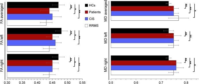
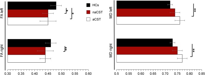
Objective: We investigated corticospinal tract (CST) integrity in the absence of white matter (WM) lesions using diffusion tensor imaging (DTI) in early MS disease stages.
Methods: Our study comprised 19 patients with clinically isolated syndrome (CIS), 11 patients with relapsing-remitting MS (RRMS), and 32 age- and sex-matched healthy controls, for whom MRI measures of CST integrity (fractional anisotropy [FA], mean diffusivity [MD]), T1- and T2-based lesion load, and brain volumes were available. The mean (SD) disease duration was 3.5 (2.1) months, and disability score was low (median Expanded Disability Status Scale 1.5) at the time of the study.
Results: Patients with CIS and RRMS had significantly lower CST FA and higher CST MD values compared with controls. These findings were present, irrespective of whether WM lesions affected the CST. However, no group differences in the overall gray or WM volume were identified.
Conclusions: In early MS disease stages, CST integrity is already affected in the absence of WM lesions or brain atrophy.
Figures

References
-
- Compston A, Coles A. Multiple sclerosis. Lancet 2008;372:1502–1517. - PubMed
-
- Barkhof F. The clinico-radiological paradox in multiple sclerosis revisited. Curr Opin Neurol 2002;15:239–245. - PubMed
-
- Trapp BD, Peterson J, Ransohoff RM, Rudick R, Mörk S, Bö L. Axonal transection in the lesions of multiple sclerosis. N Engl J Med 1998;338:278–285. - PubMed
-
- Seewann A, Vrenken H, van der Valk P, et al. . Diffusely abnormal white matter in chronic multiple sclerosis: imaging and histopathologic analysis. Arch Neurol 2009;66:601–609. - PubMed
LinkOut - more resources
Full Text Sources
Other Literature Sources