. 2017 Sep 27;2(5):e00146-17.
doi: 10.1128/mSphere.00146-17. eCollection 2017 Sep-Oct.

Comparative Analysis of Bacterial Community Composition and Structure in Clinically Symptomatic and Asymptomatic Central Venous Catheters

Affiliations

Comparative Analysis of Bacterial Community Composition and Structure in Clinically Symptomatic and Asymptomatic Central Venous Catheters

Franziska A Stressmann et al. mSphere. .

Abstract

Totally implanted venous access ports (TIVAPs) are commonly used catheters for the management of acute or chronic pathologies. Although these devices improve health care, repeated use of this type of device for venous access over long periods of time is also associated with risk of colonization and infection by pathogenic bacteria, often originating from skin. However, although the skin microbiota is composed of both pathogenic and nonpathogenic bacteria, the extent and the consequences of TIVAP colonization by nonpathogenic bacteria have rarely been studied. Here, we used culture-dependent and 16S rRNA gene-based culture-independent approaches to identify differences in bacterial colonization of TIVAPs obtained from two French hospitals. To explore the relationships between nonpathogenic organisms colonizing TIVAPs and the potential risk of infection, we analyzed the bacterial community parameters between TIVAPs suspected (symptomatic) or not (asymptomatic) of infection. Although we did not find a particular species assemblage or community marker to distinguish infection risk on an individual sample level, we identified differences in bacterial community composition, diversity, and structure between clinically symptomatic and asymptomatic TIVAPs that could be explored further. This study therefore provides a new view of bacterial communities and colonization patterns in intravascular TIVAPs and suggests that microbial ecology approaches could improve our understanding of device-associated infections and could be a prognostic tool to monitor the evolution of bacterial communities in implants and their potential susceptibility to infections. IMPORTANCE Totally implanted venous access ports (TIVAPs) are commonly used implants for the management of acute or chronic pathologies. Although their use improves the patient's health care and quality of life, they are associated with a risk of infection and subsequent clinical complications, often leading to implant removal. While all TIVAPs appear to be colonized, only a fraction become infected, and the relationship between nonpathogenic organisms colonizing TIVAPs and the potential risk of infection is unknown. We explored bacteria present on TIVAPs implanted in patients with or without signs of TIVAP infection and identified differences in phylum composition and community structure. Our data suggest that the microbial ecology of intravascular devices could be predictive of TIVAP infection status and that ultimately a microbial ecological signature could be identified as a tool to predict TIVAP infection susceptibility and improve clinical management.

Keywords: bacterial community; biofilm; catheter colonization; ecology.


Figures
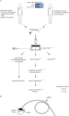
FIG 1
FIG 1
TIVAP catheter collection from patients suspected or not of catheter-related infection from two French hospitals. (A) Study design. Sterile reagents and techniques were used throughout the study, and the protocol was designed to minimize the possibilities for bacterial contamination as much as possible. Furthermore, samples of used reagents (green asterisks), materials (blue asterisks), and blanks (water used as the template in the procedure; red asterisks) as procedure controls were taken at all steps and also submitted to 16S rRNA sequencing for species identification. (B) TIVAP parts. The TIVAP is composed of a subcutaneously implanted chamber/hub (the reservoir) connected to a catheter that is inserted into a central vein (e.g., jugular or subclavian).
FIG 2
FIG 2
Heat map of species presence and their relative abundance detected in clone libraries from all 40 samples. The gray scale represents the frequency at which each species was detected in the clone library. Black arrowheads designate species generally considered of clinical origin. White arrowheads designate species of environmental origin.
FIG 3
FIG 3
Chamber septum microscopy reveals the presence of bacteria. Cell content on TIVAP septum surfaces from Limoges Hospital (after freeze-thaw treatment) was stained with nucleic acid stains SYTO9 (green, live cells) and propidium iodide (red, membrane-compromised cells). Epifluorescence and corresponding phase-contrast images of chamber septa from four symptomatic patients (A) and of chamber septa from four asymptomatic patients (B). The stain was not exclusive to bacteria; large (>5-μm) staining patterns illustrate the presence of host cell nuclei. Bar, 20 μm.
FIG 4
FIG 4
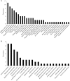
Species occurrence in all TIVAP chambers from Curie (A) and Limoges (B) Hospitals. Number of times that clones from a particular species/OTU were detected in each hospital group.
FIG 5
FIG 5
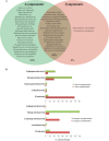
Species occurrence and phylum distribution in symptomatic and asymptomatic samples from both hospitals. (A) Species occurrence. The Venn diagram shows species (OTUs) occurring in either sample group alone or in both sample groups (brown-shaded area). Percentages indicate the number of OTUs under each condition compared to the overall total. (B) Phylum distribution.
FIG 6
FIG 6
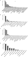
Species abundance rank order. Species/OTUs are shown ranked according to their clone library abundance in the sample group. (A) Curie asymptomatic samples; (B) Curie symptomatic samples; (C) Limoges asymptomatic samples; (D) Limoges symptomatic samples.
FIG 7
FIG 7
Slopes of the rank abundance curves for all sample groups. For each sample group, bacterial species were ranked according to their frequency in the clone library and the slope was calculated for this rank abundance curve.

References

    1. Raad I. 1998. Intravascular-catheter-related infections. Lancet 351:893–898. doi: 10.1016/S0140-6736(97)10006-X. - DOI - PubMed
    1. Raad II, Hanna HA. 2002. Intravascular catheter-related infections: new horizons and recent advances. Arch Intern Med 162:871–878. doi: 10.1001/archinte.162.8.871. - DOI - PubMed
    1. Mermel LA, Allon M, Bouza E, Craven DE, Flynn P, O’Grady NP, Raad II, Rijnders BJ, Sherertz RJ, Warren DK. 2009. Clinical practice guidelines for the diagnosis and management of intravascular catheter-related infection: 2009 update by the Infectious Diseases Society of America. Clin Infect Dis 49:1–45. doi: 10.1086/599376. - DOI - PMC - PubMed
    1. Leonidou L, Gogos CA. 2010. Catheter-related bloodstream infections: catheter management according to pathogen. Int J Antimicrob Agents 36(Suppl 2):S26–S32. doi: 10.1016/j.ijantimicag.2010.11.004. - DOI - PubMed
    1. Allegranzi B, Bagheri Nejad S, Combescure C, Graafmans W, Attar H, Donaldson L, Pittet D. 2011. Burden of endemic health care-associated infection in developing countries: systematic review and meta-analysis. Lancet 377:228–241. doi: 10.1016/S0140-6736(10)61458-4. - DOI - PubMed

LinkOut - more resources