Review
. 2018 Jan;17(1):57-78.
doi: 10.1038/nrd.2017.152. Epub 2017 Sep 29.

Deubiquitylating enzymes and drug discovery: emerging opportunities

Affiliations
Review

Deubiquitylating enzymes and drug discovery: emerging opportunities

Jeanine A Harrigan et al. Nat Rev Drug Discov. 2018 Jan.

Abstract

More than a decade after a Nobel Prize was awarded for the discovery of the ubiquitin-proteasome system and clinical approval of proteasome and ubiquitin E3 ligase inhibitors, first-generation deubiquitylating enzyme (DUB) inhibitors are now approaching clinical trials. However, although our knowledge of the physiological and pathophysiological roles of DUBs has evolved tremendously, the clinical development of selective DUB inhibitors has been challenging. In this Review, we discuss these issues and highlight recent advances in our understanding of DUB enzymology and biology as well as technological improvements that have contributed to the current interest in DUBs as therapeutic targets in diseases ranging from oncology to neurodegeneration.


Conflict of interest statement

S.P.J. is part-time employed by Mission Therapeutics, Cambridge, UK, owns shares and is a director of the company. J.A.H. is a full-time employee of Mission Therapeutics. N.M.M. and X.J. own shares in Mission Therapeutics.

Figures
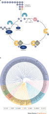
Figure 1
Figure 1. The ubiquitylation cascade and the deubiquitylase family of proteins.
a | Schematic of key events in ubiquitylation and deubiquitylation. The E1 enzyme activates ubiquitin in an ATP-dependent manner, resulting in a covalent thioester linkage between ubiquitin and the E1 cysteine residue. Ubiquitin is then transferred to an E2 conjugating enzyme, forming a thioester linkage with the catalytic cysteine. Finally, an E3 ligase assists or directly catalyses the transfer of ubiquitin from the E2 to a substrate, usually via a lysine side chain. An example of a HECT (homologous to the E6AP carboxyl terminus) or RBR (RING-between-RING) E3 ligase is shown. In subsequent rounds, ubiquitin molecules can be conjugated to the N-terminal amino group or lysines on ubiquitin itself to form chains. Deubiquitylating enzymes (DUBs) remove ubiquitin molecules from substrates or process ubiquitin precursors to generate free ubiquitin pools. b | DUB phylogenetic tree. Sequences for full-length DUB and SENP (sentrin/SUMO-specific protease) proteins were aligned with COBALT (constraint-based multiple alignment tool), a computational tool for multiple protein sequences, and subsequently visualized with FigTree v1.4.3. In regard to USP17-like, note that various related human USP17-like DUBs exist. AMSH, associated molecule with the SH3 domain of STAM; AMSHLP, AMSH-like protease; ATXN3, ataxin 3; BAP1, BRCA1-associated protein 1; CEZANNE, cellular zinc finger anti-NF-κB protein; CSN, COP9 signalosome complex subunit; CYLD, cylindromatosis; DESI, desumoylating isopeptidase; EIF3, eukaryotic translation initiation factor 3; JAMM, JAB1, MPN, MOV34 family; JOSD, josephin domain; MINDY, motif-interacting with ubiquitin-containing novel DUB family; MJD, Machado–Josephin domain-containing protease; OTUD, OTU domain-containing protein; PRPF8, pre-mRNA-processing splicing factor 8; UCHL, ubiquitin carboxy-terminal hydrolase-like; USP, ubiquitin-specific protease; VCPIP1, valosin-containing protein p97/p47 complex-interacting protein 1. PowerPoint slide
Figure 2
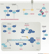
Figure 2. Various roles of DUBs in oncology.
Selected, representative examples of deubiquitylating enzymes (DUBs; light blue ovals) involved in distinct cellular pathways and regulation of various ubiquitylated substrates (dark blue ovals) related to oncology. The proteasome and associated DUBs facilitate protein turnover and recycle ubiquitin. Ubiquitin-specific protease 28 (USP28) regulates turnover of the oncogene product MYC, ataxin 3 (ATXN3) controls the stability of the tumour suppressor p53, and USP7 regulates p53 and its E3 ubiquitin ligase human double minute 2 (HDM2). USP1, USP4 and USP11 have important roles in DNA damage repair, whereas USP9X regulates claspin and is linked to replication stress and checkpoint signalling. BRCA1-associated protein 1 (BAP1) and USP22 participate in chromatin remodelling by deubiquitylating histones, and ubiquitin carboxy-terminal hydrolase-like 1 (UCHL1) plays a part in AKT signalling. These are representative examples only and not meant to be exhaustive. Examples of small-molecule compounds targeting these DUBs are shown. BRCA1, breast cancer type 1 susceptibility protein; CTIP, C-terminal-binding protein-interacting protein; FANCD2, Fanconi anaemia group D2 protein; GFR, growth factor receptor; MRE11, meiotic recombination 11 homologue 1; NBS1, Nijmegen breakage syndrome protein 1; PALB2, partner and localizer of BRCA2; PCNA, proliferating cell nuclear antigen; PI3K, phosphoinositide 3-kinase. PowerPoint slide

References

    1. Ciechanover A. The ubiquitin proteolytic system and pathogenesis of human diseases: a novel platform for mechanism-based drug targeting. Biochem. Soc. Trans. 2003;31:474–481. - PubMed
    1. Ciechanover A. Proteolysis: from the lysosome to ubiquitin and the proteasome. Nat. Rev. Mol. Cell Biol. 2005;6:79–87. - PubMed
    1. Gallastegui N, Groll M. The 26S proteasome: assembly and function of a destructive machine. Trends Biochem. Sci. 2010;35:634–642. - PubMed
    1. Finley D, Chen X, Walters KJ. Gates, channels, and switches: elements of the proteasome machine. Trends Biochem. Sci. 2016;41:77–93. - PMC - PubMed
    1. Peth A, Besche HC, Goldberg AL. Ubiquitinated proteins activate the proteasome by binding to Usp14/Ubp6, which causes 20S gate opening. Mol. Cell. 2009;36:794–804. - PMC - PubMed

Publication types

MeSH terms