. 2017 Sep 28;549(7673):533-537.
doi: 10.1038/nature24014. Epub 2017 Sep 20.

Targeting neuronal activity-regulated neuroligin-3 dependency in high-grade glioma

Affiliations

Targeting neuronal activity-regulated neuroligin-3 dependency in high-grade glioma

Humsa S Venkatesh et al. Nature. .

Abstract

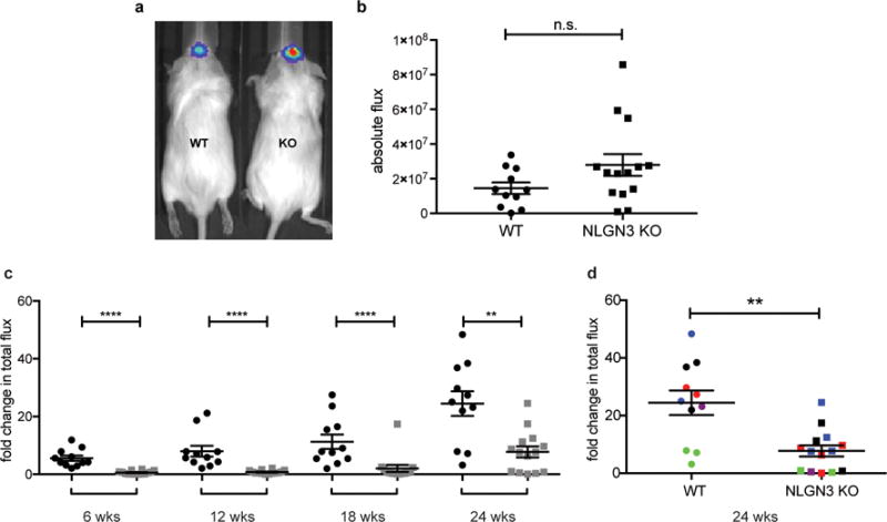
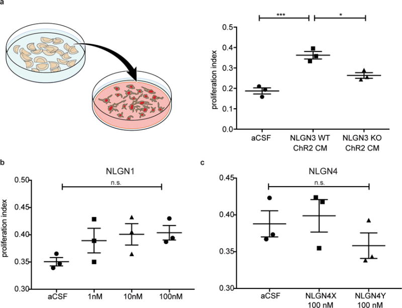
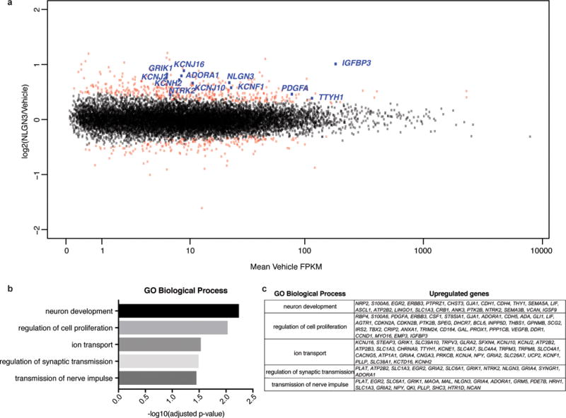
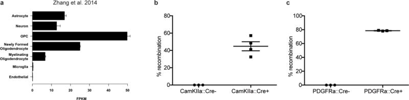
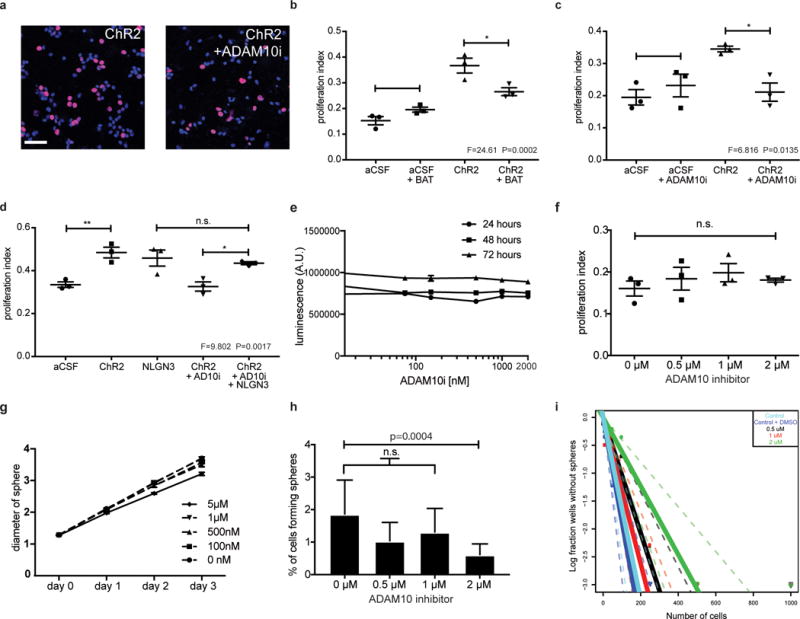
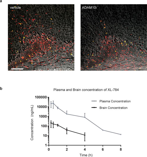
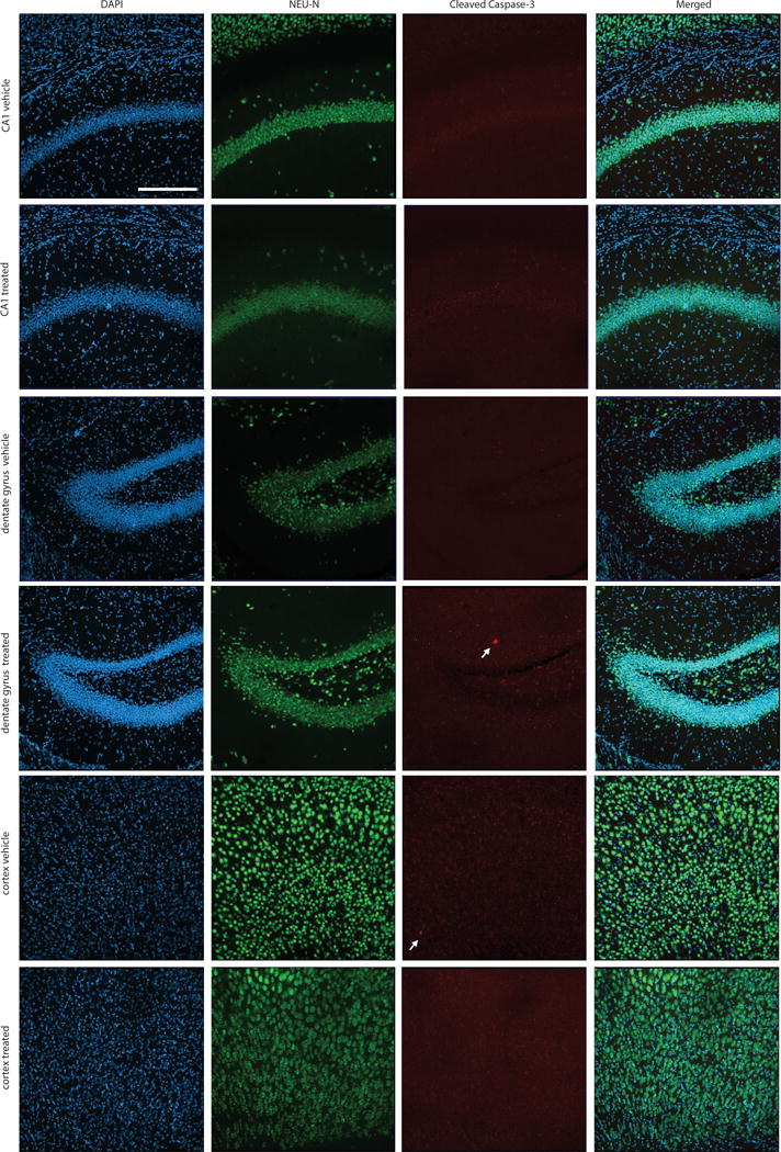
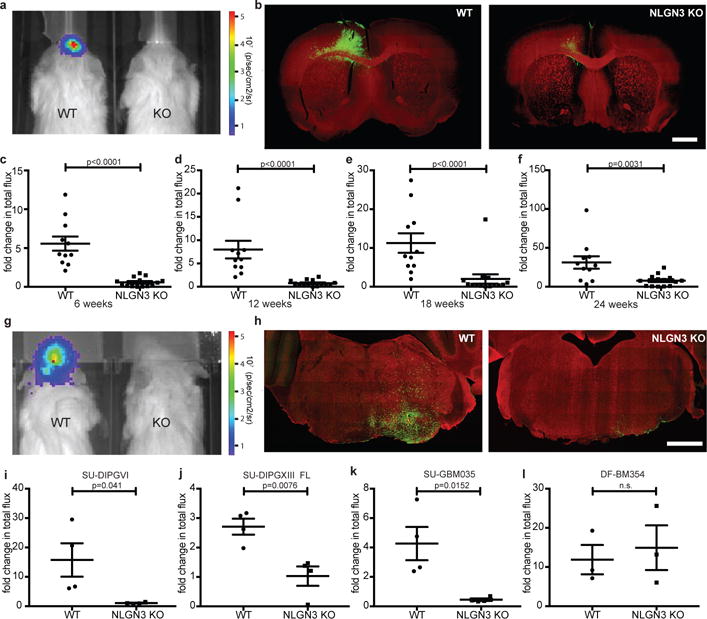
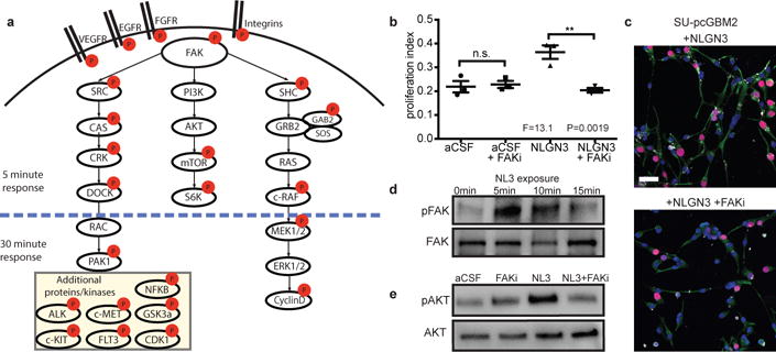
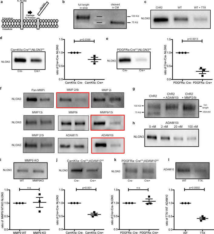
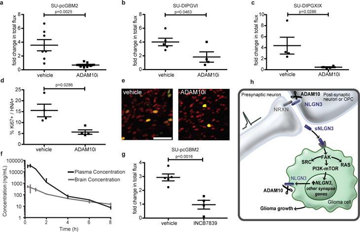
High-grade gliomas (HGG) are a devastating group of cancers, and represent the leading cause of brain tumour-related death in both children and adults. Therapies aimed at mechanisms intrinsic to glioma cells have translated to only limited success; effective therapeutic strategies will need also to target elements of the tumour microenvironment that promote glioma progression. Neuronal activity promotes the growth of a range of molecularly and clinically distinct HGG types, including adult and paediatric glioblastoma (GBM), anaplastic oligodendroglioma, and diffuse intrinsic pontine glioma (DIPG). An important mechanism that mediates this neural regulation of brain cancer is activity-dependent cleavage and secretion of the synaptic adhesion molecule neuroligin-3 (NLGN3), which promotes glioma proliferation through the PI3K-mTOR pathway. However, the necessity of NLGN3 for glioma growth, the proteolytic mechanism of NLGN3 secretion, and the further molecular consequences of NLGN3 secretion in glioma cells remain unknown. Here we show that HGG growth depends on microenvironmental NLGN3, identify signalling cascades downstream of NLGN3 binding in glioma, and determine a therapeutically targetable mechanism of secretion. Patient-derived orthotopic xenografts of paediatric GBM, DIPG and adult GBM fail to grow in Nlgn3 knockout mice. NLGN3 stimulates several oncogenic pathways, such as early focal adhesion kinase activation upstream of PI3K-mTOR, and induces transcriptional changes that include upregulation of several synapse-related genes in glioma cells. NLGN3 is cleaved from both neurons and oligodendrocyte precursor cells via the ADAM10 sheddase. ADAM10 inhibitors prevent the release of NLGN3 into the tumour microenvironment and robustly block HGG xenograft growth. This work defines a promising strategy for targeting NLGN3 secretion, which could prove transformative for HGG therapy.


Conflict of interest statement

The authors declare competing financial interests: M.M. and H.S.V. declare that Stanford University filed a patent application (15/011260) related to this work.

Figures

Extended Data Figure 1
Extended Data Figure 1. Orthotopic xenografts of pediatric glioblastoma fail to grow in the neuroligin-3-deficient brain
a, Engraftment is equivalent in Nlgn3 knockout and wild type mice. In vivo bioluminescence imaging of SU-pcGBM2 xenografts two weeks following xenograft in WT;NSG (“WT”; left) or Nlgn3 KO;NSG mice (“KO”; right). The heat map superimposed over the mouse heads represents the degree of photon emission by cells expressing firefly luciferase. b, Absolute flux of pHGG cells in identically manipulated WT;NSG (n=11) and Nlgn3 KO;NSG (n=14) mice, measured by IVIS imaging two weeks post-xenograft illustrates no significant difference in tumor engraftment. Two-sided Mann-Whitney test, n.s. = not significant (P > 0.05). Data are shown as mean +/− s.e.m. c-d, Data from main Figure 1 shown on the same axis (c) and with each independent cohort color coded for comparison of littermates (d). Data illustrate growth of pHGG (SU-pcGBM2) xenografts in identically manipulated WT;NSG (black dots, n=11) and Nlgn3y/−;NSG (grey dots, n=14) mice, measured by IVIS imaging (fold change in total photon flux) and shown at 6, 12, 18, and 24 weeks post-xenograft. Data were replicated in five independent cohorts (litters) of mice xenografted with different cell preparations on different days and the data from these five biological replicates are shown combined with each cohort color-coded (i.e. littermates are shown in the same color). **P < 0.01, ****P < 0.0001, two-sided Mann-Whitney test. Data are shown as mean +/− s.e.m.
Extended Data Figure 2
Extended Data Figure 2. Neuroligin-3 is the only neuroligin family member that promotes glioma proliferation
a, Schematic representation of active conditioned medium generation (left). Proliferation index (EdU+ and DAPI co-positive nuclei/total DAPI+ nuclei) of pHGG cells (SU-pcGBM2) exposed to plain medium (aCSF), optogenetically stimulated Nlgn3 WT cortical slice CM, or optogenetically stimulated Nlgn3 KO cortical slice CM (F = 30.8, P < 0.001). b-c, Proliferation index of patient-derived pediatric cortical glioblastoma (SU-pcGBM2) cells as measured by EdU incorporation 24 hrs after in vitro exposure to (b) recombinant human neuroligin-1 (NLGN1) at concentrations ranging from 0-100 nM or (c) recombinant human neuroligin-4X (NLGN4X) or neuroligin-4Y (NLGN4Y) at 100 nM. n=3 wells per condition. All data are presented as mean +/− s.e.m. n.s. = not significant (p > 0.05) by one-way ANOVA with Tukey’s post hoc test for multiple comparisons. *P < 0.05, ***P < 0.001.
Extended Data Figure 3
Extended Data Figure 3. Gene expression changes induced by neuroligin-3 in glioma
a, Scatterplot showing SU-pcGBM2 (n=2) gene expression changes following 16 hours of treatment with vehicle (~1% DMSO) or NLGN3 (100 nM). The x-axis shows mean FPKM value in vehicle treated cells and the y-axis shows log2(fold-change) of NLGN3 over vehicle. Points shown in red represent genes showing statistically significant change (adjusted p value < 0.1, Benjamini-Hochberg for multiple comparison testing). b, Gene Ontology Biological Processes enriched in significantly upregulated genes with NLGN3 treatment, as identified by DAVID with p values shown with Benjamini-Hochberg adjustment for multiple comparison testing. c, Genes associated with each GO BP term shown in (b).
Extended Data Figure 4
Extended Data Figure 4. Neuroligin-3 expression data and efficiency of Cre driver mice
a, Nlgn3 RNA expression (FPKM values) in various cell types; (data from Brain-seq Barres dataset) b-c, Recombination rate of inducible Cre driver models 7 days after treatment with tamoxifen (100mg/kg for 5 days) in Rosa26::tdTomatolox-stop-lox reporter mice. b, To assess the neuron-specific CamKIIa:CreER Cre driver, recombination efficiency was quantified as percent of NeuN+ neurons that co-express tdTomato+ in the cortex of either CamKIIa:CreER− or CamKIIa:CreER+ mice 7 days following completion of tamoxifen administration. c, To assess the OPC-specific Cre driver PDGFRa:CreER, recombination efficiency was quantified as number PDGFRα+ OPCs that co-express tdTomato in the cortex of either PDGFRa:CreER− or PDGFRa:CreER+ mice. n=3 mice per group.
Extended Data Figure 5
Extended Data Figure 5. NLGN3 shedding from glioma cells is regulated by NLGN3 exposure and is mediated by ADAM10
a, NLGN3 Western blot illustrating neuroligin-3 secreted into CM from optogenetically stimulated Thy1::ChR2; NSG cortical slices (ChR2 stim slice) or SU-DIPGXIII xenograft-bearing Thy1::ChR2; NSG cortical slices (ChR2 stim slice with xenograft). Performed in biological duplicate. b, NLGN3 western blot illustrating neuroligin-3 secreted into CM from wild type brain slices (WT), WT brain slices bearing xenografts of adult GBM SU-GBM035 (WT + xeno), or from Nlgn3 knockout brains bearing SU-GBM035 xenografts (Nlgn3 KO + xeno) in the absence (left 3 lanes) or presence (right 3 lanes) of 200 nM ADAM10 inhibitor GI254023X (+ADAM10i). Performed in biological triplicate. c, NLGN3 Western illustrating glioma cell secretion of NLGN3 in vitro at baseline medium conditions (aCSF), following exposure to recombinant NLGN3 with subsequent washing (NLGN3), at baseline medium conditions in the presence of ADAM10 inhibitor GI254023X (aCSF + ADAM10i) or following NLGN3 exposure in the presence of ADAM10 inhibitor (NLGN3+ADAM10i). Performed in biological triplicate. d, mRNA expression levels of ADAM10 in primary tumor and cultures of DIPG by RNA-seq with values reported as FPKM, (left; n=8 primary samples, n=7 culture samples) and in 493 individual adult glioblastoma samples from TCGA (right). Values are reported as robust multi-array averages (RMA; right). Boxes show the median, 25th and 75th percentiles, error bars show the minima and maxima.
Extended Data Figure 6
Extended Data Figure 6. Functional consequences for glioma of protease inhibition in situ and in vitro
a, SU-pcGBM2 cells (EdU, red; DAPI, blue) exposed to CM +/− ADAM10 inhibitor; Scale bar=50μm. b-d, Proliferation indices of SU-pcGBM2 cells exposed to plain medium (aCSF) or active CM +/− (b) pan-MMP inhibitor (BAT) (c) ADAM10 inhibitor or (d) ADAM10 inhibitor +/− NLGN3 rescue. n=3 wells per condition. e, Cell viability of SU-pcGBM2 cells exposed to ADAM10 inhibitor (GI254023X, 10nM-2μM) at 24-, 48-, and 72-hours, (n=3 wells/condition) f, Proliferation index of SU-pcGBM2 cells exposed to GI254023X (0-2μM; (n=3 wells/condition). g, Spheroid invasion index of SU-DIPGVI cells exposed to ADAM10i (0-5μM) at 24-, 48- and 72-hours expressed as the diameter of the sphere of glioma cells relative to the initial diameter at time 0-hours. h, Neurosphere formation assay in SU-pcGBM2 cells in the presence of GI254023X (0-2μM; n=10 wells/condition. i, Extreme limiting dilution assay (ELDA) data presented in h re-plotted here as a log fraction plot with the slope of the solid line representing the log-active cell fraction and confidence intervals shown as dotted lines. SU-pcGBM2 cells treated with ADAM10 inhibitor GI254023X at 0.5μM (black), 1μM (red) or 2μM (green), with vehicle (DMSO) control (royal blue) or no DMSO (cyan) and analyzed for neurosphere formation at two weeks. For (b-d, f), P values as indicated; one-way ANOVA with Tukey’s post hoc test for multiple comparisons; data presented as mean +/− s.e.m. For (h), χ2 test; data presented as mean +/− confidence intervals.
Extended Data Figure 7
Extended Data Figure 7. Glioma xenograft proliferation after ADAM10 inhibition and pharmacokinetics of XL-784
a, Representative confocal images (Ki67, green; human nuclear antigen, red; myelin basic protein, white) of vehicle-treated or ADAM10i-treated mice bearing frontal cortex SU-pcGBM2 xenografts; images similar to but lower magnification than those shown in Fig. 4e; n=4 vehicle, n=4 ADAM10i-treated mice. Scale bar=100μm. b, Brain tissue and plasma levels of XL-784 at various time points following a single 50 mg/kg i.p. dose in NSG mice as assessed by liquid chromatography/tandem mass spectrometry (LC-MS/MS). n=3 mice at each data point. Data are shown as mean +/− st.dev.
Extended Data Figure 8
Extended Data Figure 8. Lack of detectable neurotoxicity following treatment with INCB7839
Histological assessment of neuronal integrity was performed in mice treated with INCB7839 or vehicle control. The cortex, CA1 region of the hippocampus and dentate gyrus of the hippocampus were examined immunohistologically in the hemisphere contralateral to glioma xenografts in mice treated with INCB7839 or vehicle control. Brain sections were immunostained with NeuN (green) to mark neuronal nuclei and cleaved caspase-3 (red) to mark apoptotic cells and counterstained with DAPI (Blue). Representative sections from n=4 INCB7839-treated mice and n=4 vehicle control mice were examined. Neuronal nuclei appeared morphologically normal and non-pyknotic. Extremely few cleaved caspase-3+ cells were identified in either group; a total of one cleaved caspase-3+ cell was found in each group across all mice examined (white arrows). Scale bar = 200 μm.
Figure 1
Figure 1. Microenvironmental neuroligin-3 is necessary for HGG growth
a, IVIS of WT or Nlgn3 KO mice at 3 months. Heat map, photon emission. b, Representative coronal forebrain images of xenografts in WT (n=11) and Nlgn3 KO (n=14) mice at 6 months, as in (f); GFP+ tumor cells (green) and myelin basic protein (MBP, red). Scale bar=1 mm. c-f, Fold change in total photon flux of SU-pcGBM2 xenografts in identically manipulated WT;NSG (n=11 animals) and Nlgn3y/−;NSG (n=14 animals) mice shown at 6, 12, 18, and 24 weeks. Experiment replicated in five independent cohorts of mice and data shown combined. g, Representative IVIS images of WT (left) and Nlgn3 KO (right) mice at 6 weeks following DIPG (SU-DIPG-VI) xenografting. h, Representative confocal images at the level of the pons in Nlgn3 WT (left) and Nlgn3 KO (right) mouse brains (MBP, red) bearing DIPG xenografts (green) at 6 weeks post-xenografting; as in (i), n=4/group. Scale bar=1 mm. i-l, Fold change in photon emission in (i) SU-DIPG-VI (n=4/group), (j) SU-DIPG-XIII FL (n=4/group), (k) SU-GBM035 (n=4/group), and (l) DF-BM354 (n=3 WT, n=3 KO) xenografts in WT and Nlgn3 KO mice at 6 weeks (i,j) or 4 weeks (k,l) after xenografting. Each dot represents one mouse. P values indicated on graphs, two-sided Mann-Whitney test (c-f), Student’s two-tailed t-test (i-l). Data shown as mean+/−s.e.m. 96% CI for (c) [−6.40 to −2.81]; (d) [−7.43 to −3.63]; (e) [−15.12 to −3.80]; (f) [−30.5 to −6.65]; 95% CI for (i) [−28.61 to −0.74]; (j) [−2.73 to −0.64]; (k) [−6.60 to −1.04]; (l) [−15.93 to 22.05].
Figure 2
Figure 2. Signaling consequences of neuroligin-3 in glioma
a, Schematic illustration of signaling pathways activated following NLGN3 exposure; red circles represent proteins exhibiting increased phosphorylation. b, Proliferation index of SU-pcGBM2 cells exposed to plain medium (aCSF), aCSF+5nM FAK inhibitor (FAKi), NLGN3 (50nM), or NLGN3 (50nM)+5nM FAK inhibitor (NLGN3+FAKi), (n=3 wells per condition; P and F values indicated on graph; One-way ANOVA with Tukey’s post-hoc test for multiple comparisons; Data shown as mean+/−s.e.m.). c, Representative confocal images of SU-pcGBM2 cells as in (b) exposed to NLGN3 in the absence (top) or presence of FAK inhibitor (bottom). Vimentin, green; phospho-FAK, white; DAPI, blue; EdU, red, scale bar=50 μm. d, Representative Westerns demonstrating increased phosphorylation of FAK (Tyr861) after 0, 5, 10 or 15-minute NLGN3 exposure. e, Representative Westerns demonstrating AKTS473 phosphorylation in SU-pcGBM2 cells exposed to plain medium (aCSF), aCSF+5nM FAK inhibitor (FAKi), NLGN3 (50nM), or soluble NLGN3 (50nM)+5nM FAK inhibitor, (NLGN3+FAKi). d-e) performed in biological duplicate and technical triplicate (6 replicate Westerns). 95% CI for (b) aCSF vs. aCSF + FAKi, [−0.10 to 0.08]; aCSF vs. NLGN3, [−0.24 to −0.05]; aCSF vs. NLGN3+ FAKi, [−0.08 to 0.11]; aCSF + FAKi vs. NLGN3, [−0.23 to −0.04]; aCSF + FAKi vs. NLGN3+ FAKi, [−0.07 to 0.12]; NLGN3 vs. NLGN3+ FAKi, [0.07 to 0.25].
Figure 3
Figure 3. ADAM10 mediates activity-regulated neuroligin-3 shedding from both neurons and OPCs
a, Schematic depicting neuroligin-3 (NLGN3) cleavage. b, NLGN3 Western of slice lysate and conditioned medium (CM). c, NLGN3 Western of CM from optogenetically-stimulated Thy1::ChR2 slices or WT slices at baseline neuronal activity +/−tetrodotoxin (TTX). d, NLGN3 Western and quantification of CM from CamKIIa::CreER;Nlgn3fl/fl or Nlgn3fl/fl (no Cre) slices (n=3 animals). e, as in d from PDGFRa::CreER;Nlgn3fl/fl model (n=3 animals). f, NLGN3 Western of CM from optogenetically stimulated Thy1::ChR2 slices +/− inhibitors as indicated. g, NLGN3 Western of optogenetically stimulated Thy1::ChR2 slice homogenates +/− ADAM10 or MMP2/9 inhibitors. h, NLGN3 Western of CM from optogenetically-stimulated Thy1::ChR2 slices +/− indicated concentration of ADAM10 inhibitor. i, NLGN3 Western and quantification of WT and Mmp9−/− slice CM (n=3 animals), j-k, like i, in CamKIIa::CreER;ADAM10fl/fl (n=3 animals) (j), or PDGFRa::CreER;ADAM10fl/fl (n=3 animals) (k) models. l, ADAM10 Western and quantification of slice CM +/− TTX. Quantifications expressed as ratio of experimental/control. P values as indicated, two-tailed Student’s t-test. Data shown as mean+/−s.e.m. All Westerns performed with n=3 biologically independent samples. 95% CI for (d) [−0.48 to −0.03]; (e) [−0.93 to −0.46]; (i) [−0.49 to 0.55]; (j) [−0.56 to −0.30]; (k) [−0.18 to 0.31]; (l) [−0.70 to −0.45].
Figure 4
Figure 4. ADAM10 inhibition blocks glioma growth
a-c, Orthotopic xenograft growth (fold change in photon flux) following systemic administration of GI254023X or vehicle control for (a) SU-pcGBM2 (n=7 control, n=8 treated mice), (b) SU-DIPG-VI (n=5 control, n=4 treated mice), (c) SU-DIPG-XIX (n=4 control, n=4 treated mice) xenografts. d, In vivo proliferation index of SU-pcGBM2 cells in vehicle control and ADAM10i treated mice (n=4 mice/group). e, Representative confocal images (Ki67+, green; human nuclear antigen, HNA+ red) of SU-pcGBM2 xenografts in vehicle-treated or ADAM10i-treated mice (n=4 mice/group) as in (d). Scale bar=50μm. f, Pharmacokinetics of INCB7839; (n=3 mice/data point). g, Growth (fold change in photon flux) of SU-pcGBM2 xenografts following systemic administration of INCB7839 or vehicle control; (n=5 control, n=4 treated mice) h, Schematic summary; (neurexin = NRXN; soluble neuroligin-3 = sNLGN3). P values indicated in graphs. n.s. = not significant. Student’s two-tailed t-test (a,b,g,); two-sided Mann-Whitney (c,d). Each dot represents one mouse. Data are shown as mean +/− s.e.m except in (f) error bars are +/− st. dev. 95% CI for (a) [−4.53 to −1.22]; (b) [−4.32 to −0.05]; (g) [−2.91 to −1.04]; 97% CI for (c) [−8.63 to −1.66]; (d) [−19.39 to −2.32].

Comment in

  • Cancer: Drain the swamp to beat glioma.
    Taylor MD, Ramaswamy V. Taylor MD, et al. Nature. 2017 Sep 28;549(7673):460-461. doi: 10.1038/nature24141. Epub 2017 Sep 20. Nature. 2017. PMID: 28959957 No abstract available.
  • Anticancer therapy: Holding the thought.
    Villanueva MT. Villanueva MT. Nat Rev Drug Discov. 2017 Oct 30;16(11):751. doi: 10.1038/nrd.2017.216. Nat Rev Drug Discov. 2017. PMID: 29081517 No abstract available.

References

    1. Venkatesh HS, et al. Neuronal Activity Promotes Glioma Growth through Neuroligin-3 Secretion. Cell. 2015;161:803–16. - PMC - PubMed
    1. Varoqueaux F, et al. Neuroligins determine synapse maturation and function. Neuron. 2006;51:741–54. - PubMed
    1. Radyushkin K, et al. Neuroligin-3-deficient mice: model of a monogenic heritable form of autism with an olfactory deficit. Genes, Brain Behav. 2009;8:416–425. - PubMed
    1. Tabuchi K, et al. A neuroligin-3 mutation implicated in autism increases inhibitory synaptic transmission in mice. Science. 2007;318:71–6. - PMC - PubMed
    1. Etherton M, et al. Autism-linked neuroligin-3 R451C mutation differentially alters hippocampal and cortical synaptic function. Proc Natl Acad Sci. 2011;108:13764–13769. - PMC - PubMed

Publication types

MeSH terms