. 2017 Nov;12(21):2597-2609.
doi: 10.2217/nnm-2017-0161. Epub 2017 Sep 29.

Nanofiber-based sutures induce endogenous antimicrobial peptide

Affiliations

Nanofiber-based sutures induce endogenous antimicrobial peptide

Shixuan Chen et al. Nanomedicine (Lond). 2017 Nov.

Abstract

Aim: The aim of this study was to develop nanofiber-based sutures capable of inducing endogenous antimicrobial peptide production.

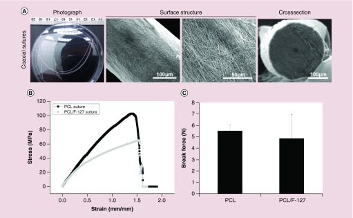
Methods: We used co-axial electrospinning deposition and rolling to fabricate sutures containing pam3CSK4 peptide and 25-hydroxyvitamin D3 (25D3).

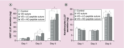
Results: The diameters and mechanical properties of the sutures were adjustable to meet the criteria of United States Pharmacopeia designation. 25D3 exhibited a sustained release from nanofiber sutures over 4 weeks. Pam3CSK4 peptide also showed an initial burst followed by a sustained release over 4 weeks. The co-delivery of 25D3 and pam3CSK4 peptide enhanced cathelicidin antimicrobial peptide production from U937 cells and keratinocytes compared with 25D3 delivery alone. In addition, the 25D3/pam3CSK4 peptide co-loaded nanofiber sutures did not significantly influence proliferation of keratinocytes, fibroblasts, or the monocytic cell lines U937 and HL-60.

Conclusion: The use of 25D3/pam3CSK4 peptide co-loaded nanofiber sutures could potentially induce endogenous antimicrobial peptide production and reduce surgical site infections.

Keywords: 25-hydroxyvitamin D3; co-delivery; electrospun nanofibers; pam3Cys-Ser-(Lys)4 peptide; surgical site infection; sutures.


Conflict of interest statement

Financial & competing interests disclosure

This work was supported partially from startup funds from University of Nebraska Medical Center, National Institute of General Medical Science (NIGMS) Grant 2P20 GM103480–06 and National Institute of General Medical Science (NIGMS) Grant R01GM123081 of the National Institutes of Health, and Otis Glebe Medical Research Foundation. The content is solely the responsibility of the authors and does not necessarily represent the official views of the National Institutes of Health. The authors have no other relevant affiliations or financial involvement with any organization or entity with a financial interest in or financial conflict with the subject matter or materials discussed in the manuscript apart from those disclosed.

No writing assistance was utilized in the production of this manuscript.

Figures

<b>Figure 1.</b>
Figure 1.. Schematic illustrating the mechanism of 25-hydroxyvitamin D3 and pam3Cys-Ser-(Lys)4 peptide co-loaded poly(ε-caprolactone) nanofiber sutures which promote antimicrobial peptide LL-37 production.
<b>Figure 2.</b>
Figure 2.. Schematic illustrating the method of fabricating nanofiber sutures.
<b>Figure 3.</b>
Figure 3.. Characterization of sutures made of random and aligned poly(ε-caprolactone) nanofibers.
(A) Photographs and scanning electron microscope images showing the morphology, surface structure and cross-section of sutures. (B) The strain–stress curves of sutures. (C) The break forces of sutures. These tests (n = 3) were repeated three times. **p < 0.01.
<b>Figure 4.</b>
Figure 4.. Characterization of sutures made of poly(ε-caprolactone) aligned nanofibers and poly(ε-caprolactone)/F-127 co-axial aligned nanofibers.
(A) Photographs and scanning electron microscope images illustrating morphology, surface structure and cross-section of sutures made of PCL/F-127 co-axial nanofibers. (B) The strain–stress curves of sutures made of PCL nanofibers and PCL/F-127 co-axial nanofibers. (C) The break forces of sutures made of PCL nanofibers and PCL/F-127 co-axial nanofibers. These tests (n = 3) were repeated three times. PCL: Poly(ε-caprolactone).
<b>Figure 5.</b>
Figure 5.. In vitro release profiles of 25-hydroxyvitamin D3 and FITC-dextran from sutures.
(A) The release profile of 25-hydroxyvitamin D3 alone loaded sutures (VD suture). (B) The release profiles of 25-hydroxyvitamin D3 and FITC-dextran (4.88 μg/ml) co-loaded sutures (VD + LC-dextran suture). (C) The release profiles of 25-hydroxyvitamin D3 and FITC-dextran (18.18 μg/ml) co-loaded sutures (VD + HC-dextran suture). These tests (n = 3) were repeated three times.
<b>Figure 6.</b>
Figure 6.. Cytotoxicity test of nanofiber sutures.
The proliferation of U937 (A), HL60 (B), keratinocytes (C) and dermal fibroblasts (D) after treatment with 1 mg/ml PCL sutures (control), 25-hydroxyvitamin D3 loaded PCL nanofiber sutures containing 100 μg/ml 25-hydroxyvitamin D3 (VD suture), 25-hydroxyvitamin D3 and peptide co-loaded PCL/pluronic F127 co-axial nanofiber sutures containing 100 μg/ml 25-hydroxyvitamin D3 and 4.88 μg/ml peptide (VD + LC-peptide suture), 25-hydroxyvitamin D3 and peptide co-loaded PCL/pluronic F127 co-axial nanofiber sutures containing 100 μg/ml 25-hydroxyvitamin D3 and 18.18 μg/ml peptide (VD + HC-peptide suture) for 1, 3 and 5 days. These tests (n = 3) were repeated three times. PCL: Poly(ε-caprolactone).
<b>Figure 7.</b>
Figure 7.. Quantification of antimicrobial peptide LL-37 expression.
The LL-37 expression of U937 cells (A) and keratinocytes (B) after treatment with 1 mg/ml PCL sutures (control), 25-hydroxyvitamin D3 loaded PCL nanofiber sutures containing 100 μg/ml 25-hydroxyvitamin D3 (VD suture), 25-hydroxyvitamin D3 and peptide co-loaded PCL/pluronic F127 co-axial nanofiber sutures containing 100 μg/ml 25-hydroxyvitamin D3 and 4.88 μg/ml peptide (VD + LC-peptide suture), 25-hydroxyvitamin D3 and peptide co-loaded PCL/pluronic F127 co-axial nanofiber sutures containing 100 μg/ml 25-hydroxyvitamin D3 and 18.18 μg/ml peptide (VD + HC-peptide suture) for 1, 3 and 5 days. These tests (n = 3) were repeated three times. PCL: Poly(ε-caprolactone).

Similar articles

Cited by

References

    1. Anderson DJ. Surgical site infections. Infect. Dis. Clin. North Am. 2011;25(1):135–153. - PubMed
    1. Hranjec T, Swenson BR, Sawyer RG. Surgical site infection prevention: how we do it. Surg. Infect. 2010;11(3):289–294. - PMC - PubMed
    1. National Institute for H and Clinical E. Surgical Site Infection: Prevention And Treatment Of Surgical Site Infection. National Institute for Health and Clinical Excellence; 2008. www.nice.org.uk/guidance/cg74
    1. Astagneau P, Rioux C, Golliot F, Brücker G. Morbidity and mortality associated with surgical site infections: results from the 1997–1999 INCISO surveillance. J. Hosp. Infect. 2001;48(4):267–274. - PubMed
    1. Justinger C, Moussavian MR, Schlueter C, et al. Antibiotic coating of abdominal closure sutures and wound infection. Surgery. 2009;145(3):330–334. - PubMed

MeSH terms

LinkOut - more resources