Review
. 2017 Nov 27:51:501-527.
doi: 10.1146/annurev-genet-120215-035312. Epub 2017 Sep 27.

Generation and Evolution of Neural Cell Types and Circuits: Insights from the Drosophila Visual System

Affiliations
Review

Generation and Evolution of Neural Cell Types and Circuits: Insights from the Drosophila Visual System

Michael Perry et al. Annu Rev Genet. .

Abstract

The Drosophila visual system has become a premier model for probing how neural diversity is generated during development. Recent work has provided deeper insight into the elaborate mechanisms that control the range of types and numbers of neurons produced, which neurons survive, and how they interact. These processes drive visual function and influence behavioral preferences. Other studies are beginning to provide insight into how neuronal diversity evolved in insects by adding new cell types and modifying neural circuits. Some of the most powerful comparisons have been those made to the Drosophila visual system, where a deeper understanding of molecular mechanisms allows for the generation of hypotheses about the evolution of neural anatomy and function. The evolution of new neural types contributes additional complexity to the brain and poses intriguing questions about how new neurons interact with existing circuitry. We explore how such individual changes in a variety of species might play a role over evolutionary timescales. Lessons learned from the fly visual system apply to other neural systems, including the fly central brain, where decisions are made and memories are stored.

Keywords: cell fate; development; evolution; neural diversity; neuropil evolution; temporal series.


Figures

Figure 1
Figure 1
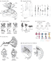
Visual system development and patterning. (a) The cell types of the retina are specified during the third larval instar. An MF sweeps across the eye imaginal disc from posterior to anterior, leaving behind evenly distributed R8 PRs. Each R8 PR then begins a recruitment process that sequentially recruits the other seven PRs in Drosophila (reviewed in 114). (b) Lamina neurons are dynamically specified one row at a time via signals from incoming PR axons (60, 61). Little is known about how the five different lamina monopolar cell types are differentially specified using similar signals from PR axons. (c) In the OPC, medulla NBs are sequentially recruited from NE and first divide to produce GMCs, which then divide to produce neurons. Each NB produces a chain of progeny. Because they were specified first, the oldest neurons appear at the bottom (81). Hth, Ey, Slp, D, and Tll are sequential transcription factors. (d) Over time, NBs change their transcriptional profiles as they transition from one transcription factor to the next in a temporal series. Loss of Ey, Slp, or D prevents transition to the next factor in the series (82). These temporal transitions help to generate much of the neural diversity of the medulla. (e) Input from regional factors across the OPC produces additional diversity (31). Together, the temporal series plus regionalization produce the highly diverse types of neurons that make up the adult medulla. (f) Specific cell types have recently been shown to form the basis of elementary motion detectors that relay information to the four layers of the lobula plate (yellow) (26, 29, 64, 65, 112). ON- (blue) and OFF- (green) edge detection relies on different pathways and cell types. (g) T4 and T5 neurons are ON- and OFF- pathway specific as well as directionally selective in the lobula plate. Shown here is R42F06–GAL4 driving CD8:GFP. It has been proposed that these neurons may be some of the most highly evolutionarily conserved cell types of the visual system (118). Abbreviations: GMC, ganglion mother cell; LPC, lamina precursor cell; MF, morphogenetic furrow; NB, neuroblast; NE, neuroepithelium; OPC, outer proliferation center; PR, photoreceptor. Panel a modified with permission from Reference , panels c and d from Reference , panel e from Reference , and panel f from Reference .
Figure 2
Figure 2
Evolution of new neural types in the visual system. (a) The eyes of female (left) and male (right) Tabanus in the family Tabanidae (the horse and deer flies) are noticeably different. The Love Spot is a specialized region of the male dorsofrontal eye found in many species of Diptera and is used for detecting and chasing females (104). (b) Some cell types in the Love Spot have become specialized for these functions. In Musca domestica, female eyes and ventral male eyes produce yellow fluorescence under blue illumination in a stochastic subset of inner R7 photoreceptors (yellow versus gray circles) (36, 51). Male retinas have specialized ommatidia in the dorsofrontal region that instead produce the same reddish fluorescence of outer, motion-sensitive photoreceptors—initially termed R7r (red circles) (36). (c) The dye-filling of photoreceptors after electrophysiological recordings was used to characterize the morphology of Musca Love Spot R7 photoreceptors (49, 52). They end in a foot in the lamina that is distinct from both inner and outer photoreceptors (49). (d) The Love Spot R7s change connectivity. Normal R7s (purple line) project to the medulla; the Love Spot R7s (red line) instead project to the lamina, like R1–6 (52). (e) Electron microscopy reconstruction of the Love Spot R7 terminals shows that they connect primarily to L2 and L3 and not to L1 (49). This suggests that they feed into the OFF pathway (green) and not ON (blue), which may reflect their function. (f) Butterflies have nine photoreceptors instead of the eight found in flies and most insects (other than Hymenoptera) (6, 105). This additional photoreceptor has been shown to be of the R7 type and is recruited in the same position as the mystery cell (M) of Drosophila, which transiently appears and disappears (105). (g) The additional R7 photoreceptor allows for the production of three stochastic outcomes in butterflies instead of the two found in Drosophila. This third stochastic type has been used to specifically deploy a newly evolved red-sensitive Rhodopsin in Papilio butterflies. (h) Evolutionary tree showing the relationships between insect orders, including the Lepidoptera and Hymenoptera (blue). Panel a modified with permission from Reference , panel b from References and , panel c from Reference , panel d from Reference , panel e from Reference , panel f from Reference , and panel h from Reference .
Figure 3
Figure 3
Diversification of insect visual systems. (a) Diagram showing morphology of Drosophila lamina cell types and the LMCs L1–L5 (33, 76, 91). (b) Honeybees have six (or more) distinct LMC morphologies (109, 110), suggesting they may have additional LMC types. (c) Lamina cell types are thought to be highly conserved across the Diptera but vary in morphology in a way that may influence connectivity and neural circuits. Here, L2 and L5 arborization patterns vary between Syrphidae and Tabanus species (17). (d) The medulla also has neurons that vary in arborization patterns across species; T5 is shown here in three families (17). These changes modify the number of columns contacted by T5 in each species. (e) Existing cell-fate specification mechanisms can be reused in new contexts in the evolution of novelty. Here, bristle specification pathways have been modified to produce butterfly wing scales, though half of the lineage undergoes targeted cell death in scales that are not innervated (39). This is strikingly similar to the use of a subset of the temporal series in the tips of the OPC, where, downstream of a Notch fate decision, half of each GMC lineage undergoes cell death to produce a more limited set of neural types (11). (f) Ornidia and Salpinogaster hoverflies (Syrphidae) produce four HS-LPTCs instead of the three found in Calliphora or Drosophila (20, 98). (g) Additional diversity can be sexually specific, as in the male-specific neurons in the Calliphora lobula plate that correspond to input from the dorsofrontal acute zone (124). Abbreviations: CBL, cell body layer; EPL, external plexiform layer; GMC, ganglion mother cell; HS-LPTCs, lobula plate tangential cells of the horizontal system; LAW, lamina area widefield; LMC, lamina monopolar cell; LVF, long visual fiber; M, medulla; MCol, male-specific columnar neurons; MLG, male-specific lobula giant tangential neurons; N, Notch; NB, neuroblast; NE, neuroepithelium; OCh, optic chiasma; OPC, outer proliferation center; SMC, sensory mother cell; S Tan, south tangential neuron; SVF, short visual fiber. Panel a modified with permission from References , , and ; panels c and d from Reference ; panel e from References and ; panel f from Reference ; and panel g from Reference . Panel b adapted from References and .
Figure 4
Figure 4
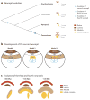
Neuropil evolution in the arthropods. (a) Phylogenetic tree of the arthropods indicating the major events in the evolution of visual system neuropils, i.e., the emergence of the second, third, and fourth neuropils. Diagrams on the right show a representative brain for each group. (b) There are three models regarding the homology of the second neuropil to the medulla or the lobula plate (LP) of extant insects. In model 1, the second neuropil is homologous to the medulla, which is generated by the outer proliferation center (OPC) (125). In this case, the inner proliferation center (IPC) also likely produces some medulla cell types. Alternatively, in model 2, the second neuropil is homologous to the LP (54), which, in extant insects, is generated by the IPC (3). Finally, in model 3, a hybrid of the extant medulla and lobula complex develops from both the OPC and IPC and may have been the second neuropil of the common ancestor of the Euarthropoda (118). (c) Given our current understanding of medulla and lobula complex development in Drosophila, the third model is the most parsimonious. In this case, chelicerates, myriapods, and branchiopods have a second neuropil that exhibits features of both the medulla and lobula complex. The first division of this neuropil generated a medulla/lobula hybrid ancestor, which was then duplicated or split into what we know today in Drosophila as the medulla and lobula.

Similar articles

Cited by

References

    1. Adolphs R. The unsolved problems of neuroscience. Trends Cogn Sci. 2015;19(4):173–75. - PMC - PubMed
    1. Ammer G, Leonhardt A, Bahl A, Dickson BJ, Borst A. Functional specialization of neural input elements to the Drosophila ON motion detector. Curr Biol. 2015;25(17):2247–53. - PubMed
    1. Apitz H, Salecker I. A region-specific neurogenesis mode requires migratory progenitors in the Drosophila visual system. Nat Neurosci. 2015;18(1):46–55. - PMC - PubMed
    1. Arendt D. The evolution of cell types in animals: emerging principles from molecular studies. Nat Rev Genet. 2008;9(11):868–82. - PubMed
    1. Arendt D, Musser JM, Baker CVH, Bergman A, Cepko C, et al. The origin and evolution of cell types. Nat Rev Genet. 2016;17(12):744–57. - PubMed

Publication types

MeSH terms

LinkOut - more resources