Review
. 2018 Feb 10:80:189-217.
doi: 10.1146/annurev-physiol-022516-034303. Epub 2017 Sep 27.

Spinal Circuits for Touch, Pain, and Itch

Affiliations
Review

Spinal Circuits for Touch, Pain, and Itch

Stephanie C Koch et al. Annu Rev Physiol. .

Abstract

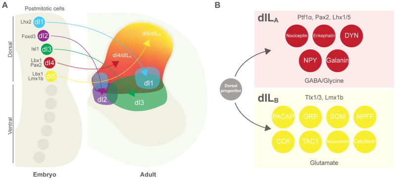
The exteroceptive somatosensory system is important for reflexive and adaptive behaviors and for the dynamic control of movement in response to external stimuli. This review outlines recent efforts using genetic approaches in the mouse to map the spinal cord circuits that transmit and gate the cutaneous somatosensory modalities of touch, pain, and itch. Recent studies have revealed an underlying modular architecture in which nociceptive, pruritic, and innocuous stimuli are processed by distinct molecularly defined interneuron cell types. These include excitatory populations that transmit information about both innocuous and painful touch and inhibitory populations that serve as a gate to prevent innocuous stimuli from activating the nociceptive and pruritic transmission pathways. By dissecting the cellular composition of dorsal-horn networks, studies are beginning to elucidate the intricate computational logic of somatosensory transformation in health and disease.

Keywords: dorsal horn; gate control; interneuron; mechanosensation; nociception; somatosensory system.


Figures

Figure 1
Figure 1. The spinal cord receives and processes information about the internal and external environment
Left: autonomic afferents monitor stretch within smooth muscle to provide information about the activity of internal organs. In the example shown, mechanoreceptors within the bladder wall activate afferent nerves as the bladder fills; in turn, a putative interneuronal pathway (dashed) within the dorsal horn mediates afferent input onto the sympathetic preganglionic nucleus (SPN), which controls bladder filling. Center: proprioceptive afferents monitor tension and stretch within striated muscle to provide information about the position of the body in space. In the example shown, Ia afferents originating in muscle spindles monitor the length and velocity of muscle fibers; in turn, Ia afferents activate agonist alpha motoneurons (MNs) as well as Ia inhibitory interneurons (INs), which suppress activity of antagonist MNs. Right: cutaneous afferents innervate the hairy and glabrous skin and are activated by mechanical, thermal, noxious or pruritic stimuli to provide information about the external environment. In the example shown, low-threshold mechanoreceptors (LTMRs) and high-threshold mechanoreceptors (HTMRs), which exhibit tuning to the intensity of mechanical stimulation of the skin, activate distinct cohorts of INs within the dorsal horn.
Figure 2
Figure 2. Neuronal diversity is established during embryonic development of the dorsal horn
A: (left) schematic cross section of the embryonic spinal cord showing transcription-factor (TF) specification and migration of dorsal horn interneurons (INs). At embryonic day 11 (E11), five early classes of postmitotic neuron (dI1–5) are present within distinct domains along the dorsoventral axis of the dorsal horn; these subsequently migrate to their positions within the mature spinal cord (right). Between E12 and E14, two further classes (dILA and dILB) are born from dorsal progenitors across a large domain occupied by dI4 and dI5 cells. B: many molecularly defined neuronal populations within the mature dorsal horn arise from late-born dILA and dILB classes: (upper panel) dILA cells are distinguished by expression of Ptf1α, Pax2 and Lhx1/5 TFs and are glycine/GABAergic; (lower panel) dILB cells are distinguished by Tlx1/3 and Lmx1b TFs and are glutamatergic. Abbreviations: CCK, cholecystokinin; DYN, dynorphin; GRP, gastrin-releasing peptide; NPY, neuropeptide Y; NPFF, neuropeptide FF; PACAP, pituitary adenylate cyclase-activating polypeptide; SOM, somatostatin; TAC1, tachykinin 1.
Figure 3
Figure 3. Molecularly defined dorsal-horn interneurons form modality-specific circuits
Composite circuit diagrams showing proposed interactions between molecularly defined classes of dorsal-horn neurons in (A) static and dynamic touch, (B) mechanical and thermal nociception, (C) itch and (D) inflammatory and neuropathic pain (see text and Tables 1 and 2 for detailed description). Dashed line: connectivity not directly demonstrated. Abbreviations: Bhlhb5, basic helix-loop-helix domain containing, class B5; CR, calretinin/calb2; DYN, dynorphin; GRP, gastrin-releasing peptide; GRPR1, gastrin-releasing peptide receptor 1; IN, interneuron; MN, motoneuron; NK1R, neurokinin 1 receptor; NPRA, natriuretic peptide receptor A; NPY, neuropeptide Y; PKCγ, protein kinase C gamma; PN, projection neuron; PV, parvalbumin; Ret, retinin; RORα, RAR-related orphan receptor alpha; SOM, somatostatin; TRPV1, transient receptor potential cation channel, subfamily V, member 1; vGluT3, vesicular glutamate transporter 3.

References

    1. Abraira VE, Ginty DD. The sensory neurons of touch. Neuron. 2013;79:618–639. - PMC - PubMed
    1. Abraira VE, Kuehn ED, Chirila AM, Springel MW, Toliver AA, Zimmerman AL, Orefice LL, Boyle KA, Bai L, Song BJ, Bashista KA, O’Neill TG, Zhuo J, Tsan C, Hoynoski J, Rutlin M, Kus L, Niederkofler V, Watanabe M, Dymecki SM, Nelson SB, Heintz N, Hughes DI, Ginty DD. The cellular and synaptic architecture of the mechanosensory dorsal horn. Cell. 2017;168:295–310. - PMC - PubMed
    1. Akay T, Tourtellotte WG, Arber S, Jessell TM. Degradation of mouse locomotor pattern in the absence of proprioceptive sensory feedback. PNAS. 2014;111:16877–16882. - PMC - PubMed
    1. Al-Khater KM, Kerr R, Todd AJ. A quantitative study of spinothalamic neurons in laminae I, III, and IV in lumbar and cervical segments of the rat spinal cord. J Comp Neurol. 2008;511:1–18. - PMC - PubMed
    1. Alba-Delgado C, El Khoueiry C, Peirs C, Dallel R, Artola A, Antri M. Subpopulations of PKCγ interneurons within the medullary dorsal horn revealed by electrophysiologic and morphologic approach. Pain. 2015;156:1714–28. - PubMed

Publication types