. 2018 Jun 1;14(6):661-667.
doi: 10.1093/ons/opx169.

A Computerized Microelectrode Recording to Magnetic Resonance Imaging Mapping System for Subthalamic Nucleus Deep Brain Stimulation Surgery

Affiliations

A Computerized Microelectrode Recording to Magnetic Resonance Imaging Mapping System for Subthalamic Nucleus Deep Brain Stimulation Surgery

Sunjay S Dodani et al. Oper Neurosurg. .

Abstract

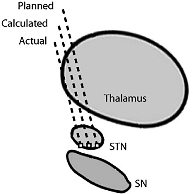
Background: Accurate electrode placement is critical to the success of deep brain stimulation (DBS) surgery. Suboptimal targeting may arise from poor initial target localization, frame-based targeting error, or intraoperative brain shift. These uncertainties can make DBS surgery challenging.

Objective: To develop a computerized system to guide subthalamic nucleus (STN) DBS electrode localization and to estimate the trajectory of intraoperative microelectrode recording (MER) on magnetic resonance (MR) images algorithmically during DBS surgery.

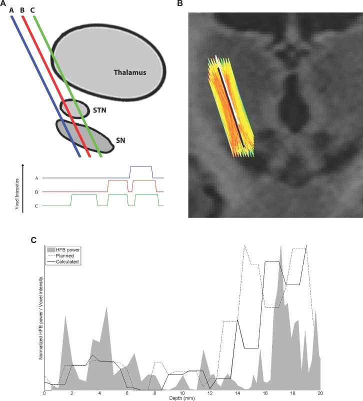
Methods: Our method is based upon the relationship between the high-frequency band (HFB; 500-2000 Hz) signal from MER and voxel intensity on MR images. The HFB profile along an MER trajectory recorded during surgery is compared to voxel intensity profiles along many potential trajectories in the region of the surgically planned trajectory. From these comparisons of HFB recordings and potential trajectories, an estimate of the MER trajectory is calculated. This calculated trajectory is then compared to actual trajectory, as estimated by postoperative high-resolution computed tomography.

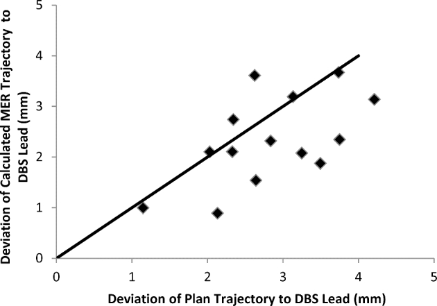
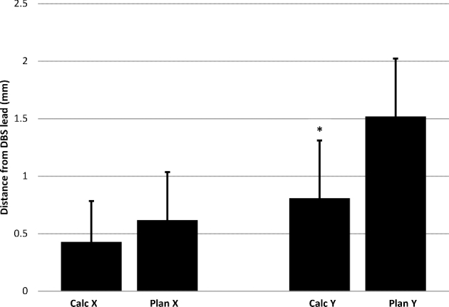
Results: We compared 20 planned, calculated, and actual trajectories in 13 patients who underwent STN DBS surgery. Targeting errors for our calculated trajectories (2.33 mm ± 0.2 mm) were significantly less than errors for surgically planned trajectories (2.83 mm ± 0.2 mm; P = .01), improving targeting prediction in 70% of individual cases (14/20). Moreover, in 4 of 4 initial MER trajectories that missed the STN, our method correctly indicated the required direction of targeting adjustment for the DBS lead to intersect the STN.

Conclusion: A computer-based algorithm simultaneously utilizing MER and MR information potentially eases electrode localization during STN DBS surgery.


Figures

FIGURE 1.
FIGURE 1.
Illustration defining trajectory terminology. “Planned” indicates the location of the surgically planned trajectory. “Actual” indicates the actual location of the DBS lead trajectory on postoperative computed tomography (CT). “Calculated” indicates the location of the intraoperative microelectrode trajectory, as calculated by the automated algorithm. The thalamus, STN, and substantia nigra structures are indicated. In this illustration, the calculated trajectory lies closer to the actual lead location than the surgical plan, indicating that the algorithm has improved lead localization estimation. SN, substantia nigra.
FIGURE 2.
FIGURE 2.
The automated algorithm. A, MR voxel intensity profile schema. Illustrative color-coded trajectories through thalamus, subthalamic nucleus, and substantia nigra are shown on an MR imaging schema (above) with corresponding color-coded voxel intensities (below). Note inversion such that positive deviations in the voxel intensity profile (below) correspond to regions of gray matter along the color-corresponding exemplar trajectory (above). B, Trajectories superimposed on 3-T coronal MR image of a patient. The surgically planned trajectory is shown in white; potential candidate trajectories used to determine the calculated trajectory are colored according to score (red: higher score, yellow: intermediate score, green: lower score). Note declining scores with distance from the planned trajectory. The final calculated trajectory is shown in black. C, Relationship of high-frequency band power to voxel intensity. HFB power (gray solid) superimposed with voxel intensity profile along the surgical plan (dashed line) and algorithm-calculated estimate (solid line) for a single trajectory. The target depth, at 15 mm, corresponds to the ventral border of STN. Noise within the data necessitates averaging of multiple, higher scoring potential trajectories to estimate the final calculated trajectory. SN, substantia nigra.
FIGURE 3.
FIGURE 3.
Determination of the calculated trajectory estimate from scored potential trajectories. The plot indicates the score distribution of 40 000 potential trajectories in the region of a single surgical plan. The top 7% of potential trajectories (scores ≥ 0.886), marked with a gray vertical line, are spatially averaged to estimate the final calculated trajectory. Due to superposition, voxel size, directional constraints, and path length constraints, some potential trajectories may superimpose.
FIGURE 4.
FIGURE 4.
Comparison of algorithm-estimated and surgically planned trajectories. Distances between surgically planned and actual trajectories (X-axis) are plotted against distances between calculated and actual trajectories (Y-axis). Points below the solid line represent cases in which the accuracy of the algorithm exceeds that of the surgical plan, while points above the line represent the opposite.
FIGURE 5.
FIGURE 5.
Comparison of algorithm-estimated and surgically planned trajectories in the axial plane of the target z-coordinate. Distances to the actual DBS lead trajectory are shown for calculated trajectories (Calc) and the surgically planned trajectories (Plan), in the mediolateral (X), and anteroposterior (Y) dimensions. Note the proportionate improvements in accuracy that are apparent for both X- and Y-coordinates, although only Y-axis improvements were statistically significant.

Similar articles

References

    1. Benabid AL, Chabardes S, Mitrofanis J, Pollak P. Deep brain stimulation of the subthalamic nucleus for the treatment of Parkinson's disease. Lancet Neurol. 2009;8(1):67-81. - PubMed
    1. Krack P, Benazzouz A, Pollak Pet al. Treatment of tremor in Parkinson's disease by subthalamic nucleus stimulation. Mov Disord. 1998;13(6):907-914. - PubMed
    1. Benabid AL, Pollak P, Gross Cet al. Acute and long-term effects of subthalamic nucleus stimulation in Parkinson's disease. Stereotact Funct Neurosurg. 1994;62(1-4):76-84. - PubMed
    1. Krack P, Pollak P, Limousin P, Benazzouz A, Benabid AL. Stimulation of subthalamic nucleus alleviates tremor in Parkinson's disease. Lancet. 1997;350(9092):1675. - PubMed
    1. Kumar R, Lozano AM, Kim YJet al. Double-blind evaluation of subthalamic nucleus deep brain stimulation in advanced Parkinson's disease. Neurology. 1998;51(3):850-855. - PubMed

Publication types