. 2017 Sep;14(3):1929-1934.
doi: 10.3892/etm.2017.4764. Epub 2017 Jul 10.

Construction and assessment of bio-engineered intervertebral discs

Affiliations

Construction and assessment of bio-engineered intervertebral discs

Hongfei Xiang et al. Exp Ther Med. 2017 Sep.

Abstract

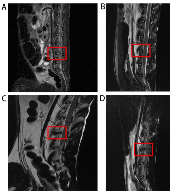
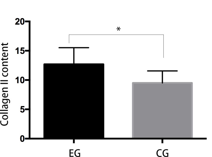
The present study assessed the value of bone marrow-mesenchymal stem cells (BM-MSCs) transformed by nucleus pulposus cells (NPs) for engineering of intervertebral discs. BM-MSCs and fetal NPs were cultured, planted onto polylactic acid-polyglycolic acid co-polymer (PLGA) and observed under inverted and scanning electron microscopes. PLGA scaffolds with adherent or suspended BM-MSCs and NPs were implanted into intervertebral discs of New Zealand white rabbits. Intervertebral signal intensity was evaluated by Thompson grading after 12 weeks. Proteoglycan and type II collagen were measured spectrophotometrically and immunohistochemically, respectively. Spindle or multi-angular BM-MSCs developed fibro-like phenotypesin co-culture with NPs and grew with a normal morphology when attached to PLGA scaffolds. A significant difference was observed in intervertebral proteoglycan expression and collagen II expression in the PLGA scaffold group vs. that in the control group implanted with BM-MSCs and NPs without a scaffold (3.93±0.31 vs. 3.52±0.26 mg/100 mg, 12.70±2.83 vs. 9.50±2.06, respectively). Thus, BM-MSCs can be co-cultured with NPs to enhance their differentiation into NPs for disc regeneration. In conclusion, PLGA scaffolds offer viable growing conditions and allow for the maintenance of mechanical properties and spatial structures of the engineered tissue, which meets the requirements of tissue-engineered discs that do not degenerate.

Keywords: interverbebral disc; mesenchymal stem cells; polylactic acid-polyglycolic acid copolymer; tissue engineering.


Figures

Figure 1.
Figure 1.
Nucleus pulposus cells were polygonal, round and fusiform, and certain cells were mitotic with a high proliferative capacity as observed under an inverted phase contrast microscope (magnification, ×400).
Figure 2.
Figure 2.
BM-MSCs had extended pseudopodia and were adherent and in an active proliferating state. Cells had fusiform, polygonal and irregular shapes as observed under an inverted phase contrast microscope (magnification, ×400). The BM-MSCs contained a large number of fluorescent particles. BM-MSCs, bone marrow-mesenchymal stem cells.
Figure 3.
Figure 3.
Mixed bone marrow-mesenchymal stem cells and nucleus pulposus cells were adherent and grew well, secreting abundant extracellular matrix that strongly fluoresced under a fluorescent microscope (magnification, ×400).
Figure 4.
Figure 4.
PLGA scaffolds had a uniform fiber mesh aperture as observed under a scanning electron microscope and cells were adherent and grew well, extending pseudopodia and secreting an abundant extracellular matrix when cells were attached and cross-linked to PLGA scaffolds. PLGA, polylactic acid-polyglycolic acid co-polymer (magnification, ×800).
Figure 5.
Figure 5.
Magnetic resonance imaging detection in the control and experimental group. (A) Control group and (B) experimental group immediately after surgery. (C) Control group and (D) experimental group after 12 weeks. The signal in the experimental group was higher than the control group. Surgery position is marked by the red frame.
Figure 6.
Figure 6.
Green fluorescence was observed in intervertebral disc slices 12 weeks after implantation, indicating bone marrow-mesenchymal stem cell survival (magnification, 400).
Figure 7.
Figure 7.
Proteoglycan content were detected by benzene-3-phenol spectrophotometric detection. EG, experimental group; CG, control group. Proteoglycan content of EG was significantly higher than that in the control group. *P<0.05.
Figure 8.
Figure 8.
Immunohistochemical detection of collagen II expression in cytoplasts in the (A) control and (B) experimental groups. Immunoreactive cells were brown or pale yellow and cells were counterstained with hematoxylin (magnification, ×400).
Figure 9.
Figure 9.
Expression of collagen II was scored according to standard methods. EG, experimental group; CG, control group. Collagen II content was significantly different between experimental and control groups. *P<0.05.

References

    1. Hoy D, Brooks P, Blyth F, Buchbinder R. The epidemiology of low back pain. Best Pract Res Clin Rheumatol. 2010;24:769–781. doi: 10.1016/j.berh.2010.10.002. - DOI - PubMed
    1. Takahashi K, Aoki Y, Ohtori S. Resolving discogenic pain. Eur Spine J. 2008;17:S428–S431. doi: 10.1007/s00586-008-0752-4. (Suppl 4) - DOI - PMC - PubMed
    1. McMeeken J, Tully E, Stillman B, Nattrass C, Bygott IL, Story I. The experience of back pain in young Australians. Man Ther. 2001;6:213–220. doi: 10.1054/math.2001.0410. - DOI - PubMed
    1. Smith LJ, Nerurkar NL, Choi KS, Harfe BD, Elliott DM. Degeneration and regeneration of the intervertebral disc: Lessons from development. Dis Model Mech. 2011;4:31–41. doi: 10.1242/dmm.006403. - DOI - PMC - PubMed
    1. Chadderdon RC, Shimer AL, Gilbertson LG, Kang JD. Advances in gene therapy for intervertebral disc degeneration. Spine J. 2004;4:S341–S347. doi: 10.1016/j.spinee.2004.07.027. (Suppl 6) - DOI - PubMed