

Silica nanoparticles induce endoplasmic reticulum stress response, oxidative stress and activate the mitogen-activated protein kinase (MAPK) signaling pathway
- PMID: 28962324
- PMCID: PMC5598250
- DOI: 10.1016/j.toxrep.2014.10.023
Silica nanoparticles induce endoplasmic reticulum stress response, oxidative stress and activate the mitogen-activated protein kinase (MAPK) signaling pathway
Abstract
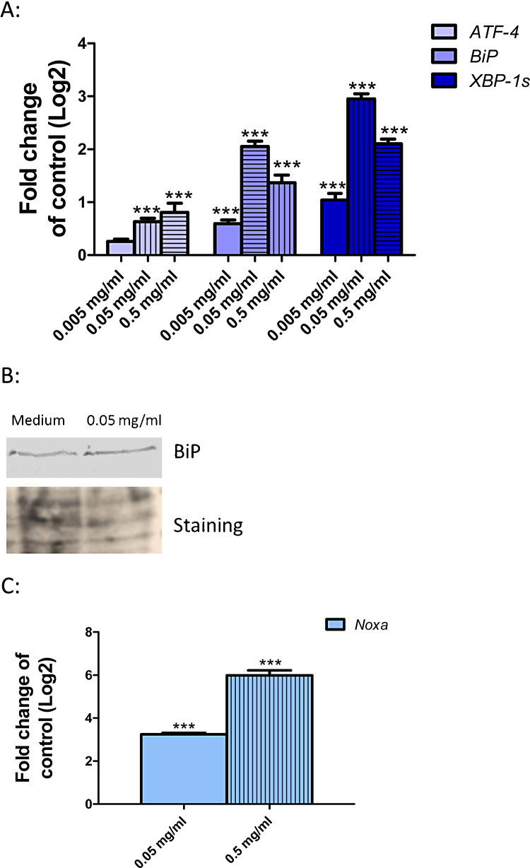
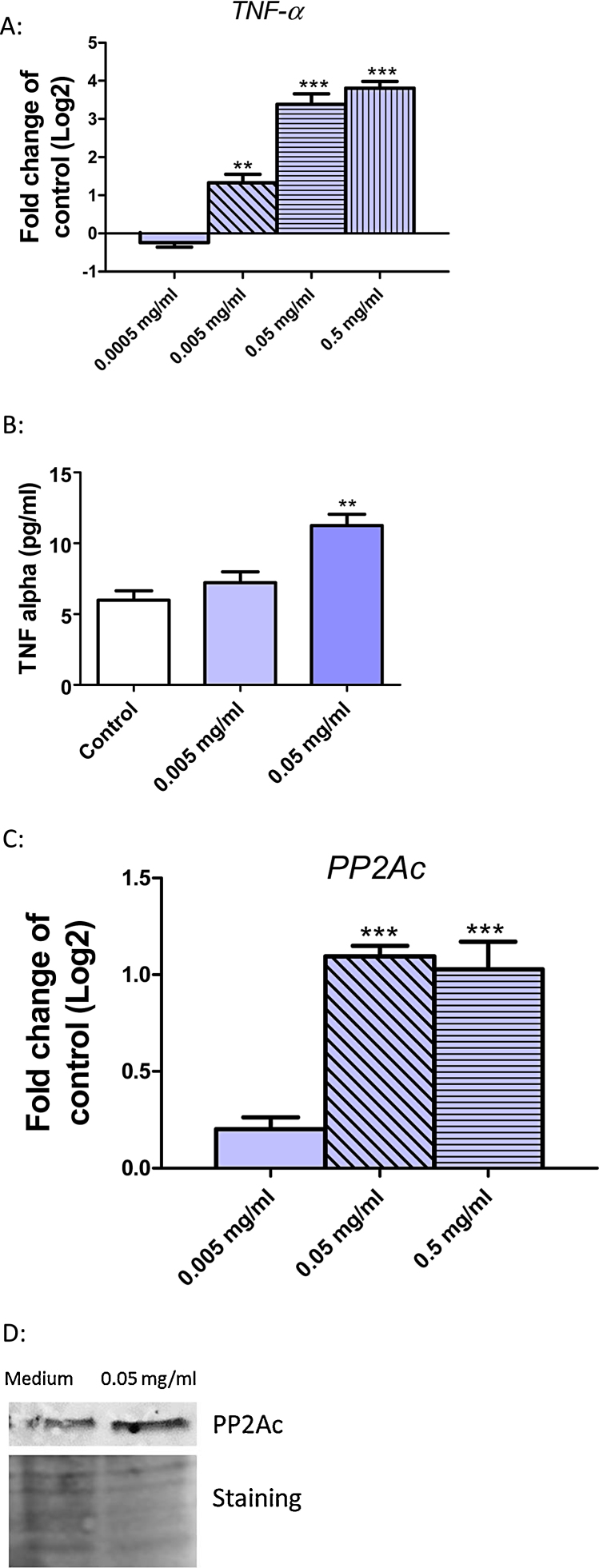
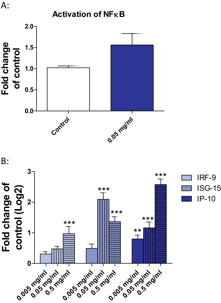
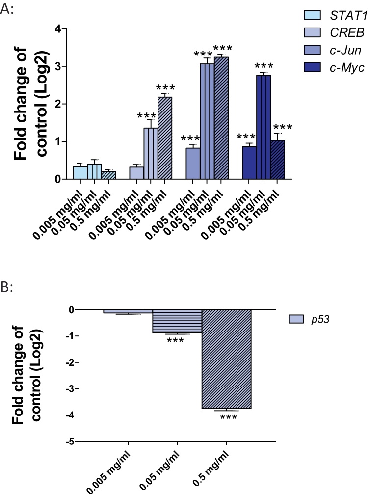
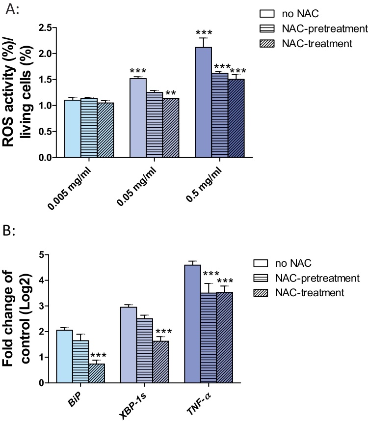
Application of silica nanoparticles (SiO2-NPs) may result in human exposure. Here we investigate unexplored modes of action by which SiO2-NPs with average size of 225 nm act on human hepatoma cells (Huh7). We focused on the endoplasmic (ER) stress response and on mitogen-activated protein kinase (MAPK) signaling pathways. Both pathways were induced. ER stress and the associated three unfolded protein response (UPR) pathways were activated as demonstrated by significant inductions of BiP and XBP-1s and a moderate but significant induction of ATF-4 at 0.05 and 0.5 mg/ml. In addition to activation of NFкB interferon stimulated genes IP-10, IRF-9, and ISG-15 were up-regulated. As a consequence of ER stress, the pro-inflammatory cytokine TNFα and PP2Ac were induced following exposure to 0.05 mg/ml SiO2-NPs. Additionally, this occurred at 0.005 mg/ml SiO2-NPs for TNFα at 24 h. This in turn led to a strong transcriptional induction of MAP-kinases and its target genes cJun, cMyc and CREB. A strong transcriptional down-regulation of the proapoptotic gene p53 occurred at 0.05 and 0.5 mg/ml SiO2-NP. Exposure of Huh7 cells to the anti-oxidant N-acetyl cysteine reduced transcriptional induction of ER stress markers demonstrating a link between the induction of oxidative stress and ER stress. Our study demonstrates that SiO2-NPs lead to strong ER stress and UPR induction, oxidative stress, activation of MAPK signaling and down-regulation of p53. All of these activated pathways, which are analyzed here for the first time in detail, inhibit apoptosis and induce cell proliferation, which may contribute to a hepatotoxic, inflammatory and tumorigenic action of SiO2-NPs.
Keywords: ATF-4, Activating transcription factor 4; ATF-6, activating transcription factor 6; BiP, binding immunoglobulin protein; CHOP, CCAAT/enhancer binding protein-homologous protein; CREB, cAMP response element-binding protein; Huh7, human hepatoma cells; Human hepatoma cells; IFN α, interferon α; IFN β, interferon β; IP-10, interferon gamma-induced protein 10; IRE-1, inositol-requiring protein 1; IRF-9, interferon regulatory factor 9; ISG-15, interferon-induced 17 kDa protein; ISGs, interferon stiulated genes; MAPK, mitogen-activated protein kinase signaling pathway; NFκB, nuclear factor ‘kappa-light-chain-enhancer’ of activated B-cells; Noxa, phorbol-12-myristate-13-acetate-induced protein 1; PERK, protein kinase like ER kinase; PP2A, protein phosphatase 2a; Proinflammatory response ;Iinterferon-stimulated genes; STAT1, signal transducer and activator of transcription 1; SiO2-NPs, silica nanoparticles; TNFα, tumor necrosis factor α; Tumor necrosis factor alpha; UPR, unfolded protein response; XBP-1, X-box binding protein 1; eIF2α, eukaryotic initiation factor 2α; p53, TP53-tumorsuppressor-gene.
Figures
References
-
- Liu T., Li L., Teng X., Huang X., Liu H., Chen D., Ren J., He J., Tang F. Single and repeated dose toxicity of mesoporous hollow silica nanoparticles in intravenously exposed mice. Biomat. 2011;32:1657–1668. - PubMed
-
- Cho M., Cho W.S., Choi M., Kim S.J., Han B.S., Kim S.H., Kim H.O., Sheen Y.Y., Jeong J. The impact of size on tissue distribution and elimination by single intravenous injection of silica nanoparticles. Toxicol. Lett. 2009;189:177–183. - PubMed
-
- Ariano P., Zamburlin P., Gilardino A., Mortera R., Onida B., Tomatis M., Ghiazza M., Fubini B., Lovisolo D. Interaction of spherical silica nanoparticles with neuronal cells: size-dependent toxicity and perturbation of calcium homeostasis. Small. 2011;7:766–774. - PubMed
-
- Christen V., Fent K. Silica nanoparticles and silver-doped silica nanoparticles induce en-doplasmatic reticulum stress response and alter cytochrome P4501A activity. Chemosphere. 2012;87:423–434. - PubMed
LinkOut - more resources
Full Text Sources
Other Literature Sources
Research Materials
Miscellaneous
