

Reduced kidney levels of lysophosphatidic acids in rats after chronic administration of aristolochic acid: Its possible protective role in renal fibrosis
- PMID: 28962344
- PMCID: PMC5598376
- DOI: 10.1016/j.toxrep.2015.02.012
Reduced kidney levels of lysophosphatidic acids in rats after chronic administration of aristolochic acid: Its possible protective role in renal fibrosis
Abstract
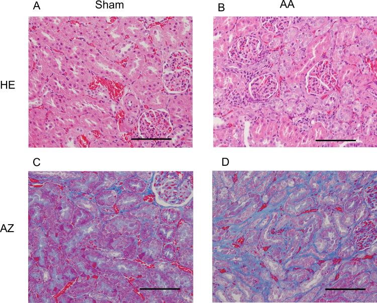
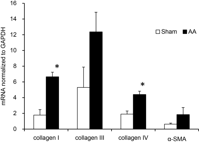
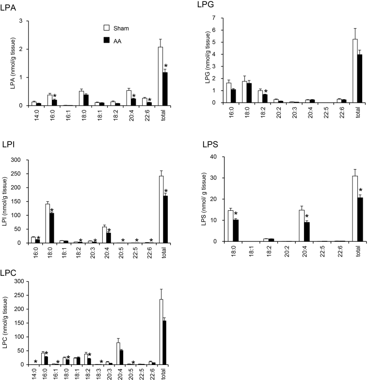
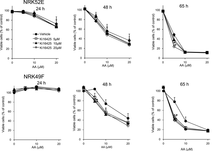
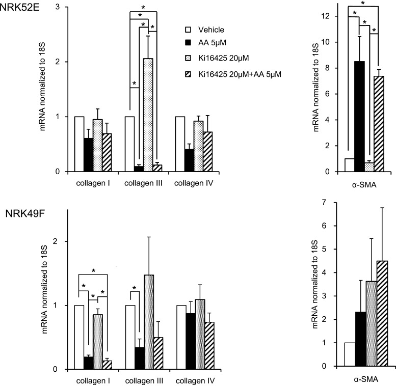
Aristolochic acid (AA) is considered to be a causative agent for progressive interstitial renal fibrosis, leading to AA nephropathy. Lysophosphatidic acid (LPA) is a mediator in the onset of renal fibrosis. In this study, we analyzed the molecular species of LPA and its precursor lysophospholipids in kidney tissue from rats exposed to AA. Daily intraperitoneal injections of AA for 35 days to rats gave rise to fibrosis in kidney, decreased the kidney levels of LPA, lysophosphatidylserine and lysophosphatidylinositol. In rat renal cell lines (NRK52E and NRK49F), AA-induced cytotoxicity was potentiated by Ki16425, LPA1,3 receptor antagonist. The level of mRNA encording α-smooth muscle actin was significantly increased by AA-treatment only in NRK52E cells, while the mRNA level of collagen III was decreased in both NRK52E and NRK49F cells. These results suggest that endogenous LPA in rat kidney prevents AA-induced renal fibrosis.
Keywords: 18S, ribosomal protein S18; AA, aristolochic acid; AZ, azan Mallory; Aristolochic acid; Chronic kidney disease; Fibrosis; GAPDH, glyceraldehyde 3-phosphate dehydrogenase; HE, hematoxylin/eosin; LC–MS/MS, liquid chromatography–tandem mass spectrometry; LPA, lysophosphatidic acid; LPC, lysophosphatidylcholine; LPE, lysophosphatidylethanolamine; LPG, lysophosphatidylglycerol; LPI, lysophosphatidylinositol; LPL, lysophospholipid; LPS, lysophosphatidylserine; Lysophosphatidic acid; Lysophospholipid; Nephrotoxicity; PLA1, phospholipase A1; PLA2, phospholipase A2; lysoPLD, lysophospholipase D; α-SMA, α-smooth muscle actin.
Figures
References
-
- Grzelczyk A., Gendaszewska-Darmach E. Novel bioactive glycerol-based lysophospholipids: new data – new insight into their function. Biochimie. 2013;95:667–679. - PubMed
-
- Makide K., Kitamura H., Sato Y., Okutani M., Aoki J. Emerging lysophospholipid mediators, lysophosphatidylserine, lysophosphatidylthreonine, lysophosphatidylethanolamine and lysophosphatidylglycerol. Prostaglandins Other Lipid Mediat. 2009;89:135–139. - PubMed
-
- Tokumura A. Metabolic pathways and physiological and pathological significances of lysolipid phosphate mediators. J. Cell. Biochem. 2004;92:869–881. - PubMed
-
- Murph M., Tanaka T., Pang J., Felix E., Liu S., Trost R., Godwin A.K., Newman R., Mills G. Liquid chromatography mass spectrometry for quantifying plasma lysophospholipids: potential biomarkers for cancer diagnosis. Methods Enzymol. 2007;433:1–25. - PubMed
-
- Tsutsumi T., Adachi M., Nikawadori M., Morishige J., Tokumura A. Presence of bioactive lysophosphatidic acid in renal effluent of rats with unilateral ureteral obstruction. Life Sci. 2011;89:195–203. - PubMed
LinkOut - more resources
Full Text Sources
Other Literature Sources
Research Materials
Miscellaneous
