. 2015 Jun 15:2:908-916.
doi: 10.1016/j.toxrep.2015.06.006. eCollection 2015.

Ascorbic acid attenuates antineoplastic drug 5-fluorouracil induced gastrointestinal toxicity in rats by modulating the expression of inflammatory mediators

Affiliations

Ascorbic acid attenuates antineoplastic drug 5-fluorouracil induced gastrointestinal toxicity in rats by modulating the expression of inflammatory mediators

Abdulrahman Khazim Al-Asmari et al. Toxicol Rep. .

Abstract

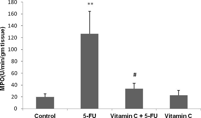
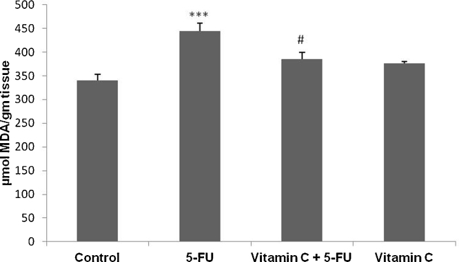
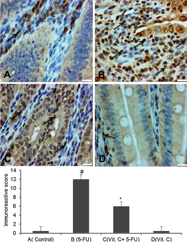
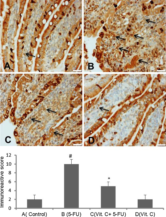
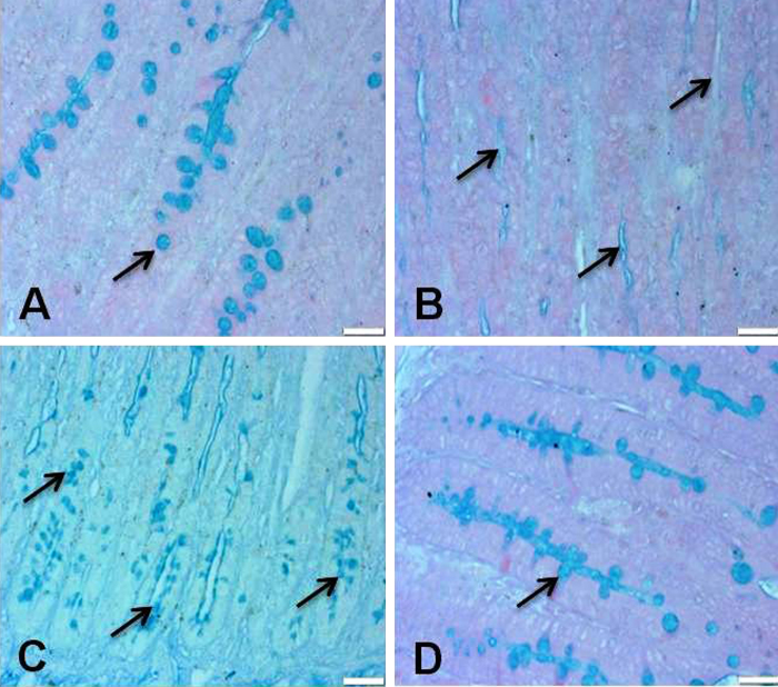
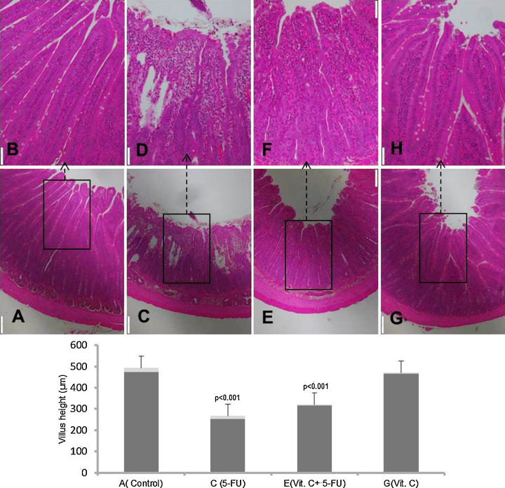
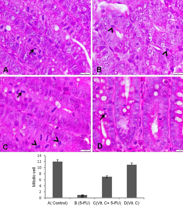
Damage to the mucous membrane is a serious issue associated with chemotherapy. Gastrointestinal (GI) toxicity is complex and multistep process and unregulated production of reactive oxygen species (ROS) and inflammatory mediators play vital role in the development of GI toxicity. In the present study we have investigated the attenuating potential of vitamin C (vit. C) on 5 fluorouracil (5-FU) induced GI toxicity by targeting oxidative stress and inflammatory markers in Sprague Dawley (SD) rats. Rats were gavaged with vit. C (500 mg/kg b. wt.) or vehicle daily (day 1-10) and were given intraperitoneal injection of 5-FU (150 mg/kg b. wt.) or saline (control) on day 8 to induce mucositis. We found that vit. C supplementation attenuated 5-FU induced lipid peroxidation, myeloperoxidase (MPO) activity, activation of NF-kB and expression of COX-2. Histological observations further supported the protective potential of vit. C against 5-FU induced intestinal anomalies such as neutrophil infiltration, loss of cellular integrity, villus and crypt deformities. Thus the biochemical, molecular and histological findings of the present study demonstrate that oxidative stress and inflammation play vital role in 5-FU induced GI toxicity and the inhibitory potential of vit. C is may be due to the modulation of oxidative stress, activation of redox sensitive transcription factor and also its downstream target molecules.

Keywords: Antineoplastic drugs; Intestinal toxicity; Oxidative stress and inflammation.


Figures

Fig. 1
Fig. 1
Effect of vit. C and 5-FU on MPO activity of small intestine. Bars represent the mean ± SEM per treatment group (= 6). **p < 0.001 shows significant difference in the animals treated with only 5-FU as compared with vehicle treated control animals. #p < 0.05, shows the statistically significant difference in the animals treated with vit. C along with 5-FU when compared with the animals injected with 5-FU only.
Fig. 2
Fig. 2
Effect of vit. C and 5-FU on MDA level of small intestine. Bars represent the mean ± SEM per treatment group (= 6). ***p < 0.001 shows significant difference in only 5-FU treated animals when compared with vehicle treated animals. #p < 0.05, shows the statistically significant difference in the animals treated with vit. C along with 5-FU when compared with the animals injected with 5-FU only.
Fig. 3
Fig. 3
Effect of vit. C on 5-FU induced NF-kB activation in intestine. Representative photomicrographs (magnification 40×; scale bar-100 μm) depicting immunohistochemical activation of NF-kB, (A) vehicle treated animals showing minimal or no staining, (B) 5-FU treated animals showing remarkably high intense staining as shown by arrows, (C) administration of vit. C + 5-FU treated animals showing moderate staining (arrows) and (D) vit. C treated animals showing minimal or no staining. Brown color indicates immunopositive staining of NF-kB and blue color indicates haematoxylin staining. (For interpretation of the references to color in this figure legend, the reader is referred to the web version of this article.)
Fig. 4
Fig. 4
Effect of vit. C on 5-FU induced expression of COX-2 in small intestine. Representative photomicrographs (magnification 20×; scale bar-100 μm)) depicting immunohistochemical activation of COX-2, (A) vehicle treated animals showing minimal or no immunopositive staining, (B) 5-FU treated animals showing remarkable intense staining as shown by arrows, (C) Vit. C + 5-FU treated animals showing low staining (arrow) and (D) Vit. C only treated animals showing minimal or no staining. Brown color indicates immunopositive staining of COX-2 and blue color depicts haematoxylin staining. (For interpretation of the references to color in this figure legend, the reader is referred to the web version of this article.)
Fig. 5
Fig. 5
Effect of vit. C treatment on 5-FU induced mucin depletion in small intestine. Representative photomicrographs (magnification 20×; scale bar-100 μm) depicting mucin staining, (A) vehicle treated animals showing normal mucin layer (arrows), (B) 5-FU treated animals showing complete mucin depletion as shown by arrows, (C) Vit. C + 5-FU treated animals showing moderate mucin depletion (arrows) and (D) Vit. C only treated animals showing normal mucin layer.
Fig. 6
Fig. 6
Effect of vit. C treatment on 5-FU induced histological alterations in small intestine. Representative photomicrographs (magnification 4× & 10×; scale bar-100 μm) depicting histology analysis, (A) only vehicle treated animals showing normal histology, (C) 5-FU treated animals showing marked histological deformities, (E) Vit. C + 5-FU treated animals showing suppressed histological alterations and (G) Vit. C only treated animals showing normal histology. Insets (B, D, F & H) at the upper panel show a magnified view (10× magnifications) of the insets (A, C, E & G) showed at the lower panel (4× magnifications).
Fig. 7
Fig. 7
Showing the mitotic and apoptotic bodies of the H & E stained small intestine crypts (magnification 40×, scale bar 100 μm). (A), vehicle treated animals showing normal mitotic cells (arrow), (B) 5-FU treated animals depicting no intact mitotic cells and number of apoptotic bodies (arrow head), (C) Vit. C + 5-FU treated animals showing renewal of mitotic cells (arrow) and suppressed apoptotic bodies (arrow head) and (D) only Vit. C treated animals showing normal mitotic cells with no apoptotic bodies.

References

    1. Bradley P.P., Priebat D.A., Christensen R.D., Rothstein G. Measurement of cutaneous inflammation: estimation of neutrophil content with an enzyme marker. J. Invest. Dermatol. 1982;78:206–209. - PubMed
    1. Cárcamo J.M., Pedraza A., Bórquez-Ojeda O., Golde D.W. Vitamin C suppresses TNFα-induced NFB activation by inhibiting I(Bα phosphorylation. Biochemistry. 2002;41:12995–13002. - PubMed
    1. Chambial S., Dwivedi S., Shukla K.K., John P.J., Sharma P. Vitamin C in disease prevention and cure: an overview. Indian J. Clin. Biochem. 2013;28:314–328. - PMC - PubMed
    1. Chang C.-T., Ho T.-Y., Lin H., Liang J.-A., Huang H.-C., Li C.-C., Lo H.-Y., Wu S.-L., Huang Y.-F., Hsiang C.-Y. 5-Fluorouracil induced intestinal mucositis via nuclear factor-(B ctivation by transcriptomic analysis and in vivo bioluminescence imaging. PLoS One. 2012;7:e31808. - PMC - PubMed
    1. Chen Y., Luo G., Yuan J., Wang Y., Yang X., Wang X., Li G., Liu Z., Zhong N. Vitamin C mitigates oxidative stress and tumor necrosis factor-alpha in severe community-acquired pneumonia and LPS-induced macrophages. Mediators Inflamm. 2014;2014:426740. - PMC - PubMed