

Glycolaldehyde induces endoplasmic reticulum stress and apoptosis in Schwann cells
- PMID: 28962488
- PMCID: PMC5598486
- DOI: 10.1016/j.toxrep.2015.10.014
Glycolaldehyde induces endoplasmic reticulum stress and apoptosis in Schwann cells
Abstract
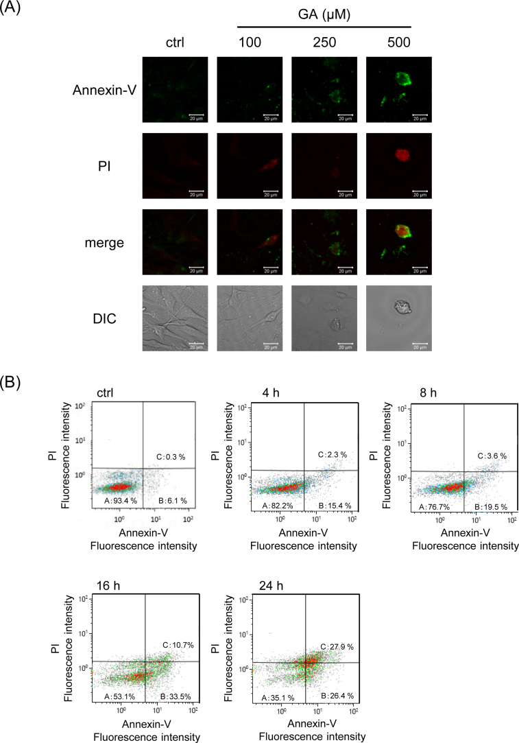
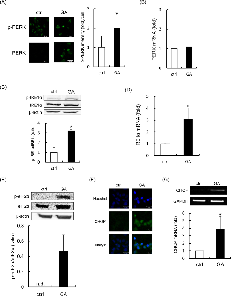
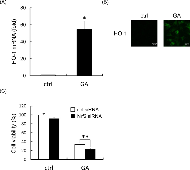
Schwann cell injury is caused by diabetic neuropathy. The apoptosis of Schwann cells plays a pivotal role in diabetic nerve dysfunction. Glycolaldehyde is a precursor of advanced glycation end products that contribute to the pathogenesis of diabetic neuropathy. In this study, we examined whether glycolaldehyde induces endoplasmic reticulum (ER) stress and apoptosis in rat Schwann cells. Schwann cells treated with 500 μM glycolaldehyde showed morphological changes characteristic of apoptosis. Glycolaldehyde activated apoptotic signals, such as caspase-3 and caspase-8. Furthermore, it induced ER stress response involving RNA-dependent protein kinase-like ER kinase (PERK), inositol-requiring ER-to-nucleus signal kinase 1α (IRE1α), and eukaryotic initiation factor 2α (eIF2α). In addition, glycolaldehyde activated CCAAT/enhancer-binding homologous protein (CHOP), an ER stress response factor crucial to executing apoptosis. Knockdown of nuclear factor E2-related factor 2 (Nrf2), which is involved in the promotion of cell survival following ER stress, enhanced glycolaldehyde-induced cytotoxicity, indicating that Nrf2 plays a protective role in the cytotoxicity caused by glycolaldehyde. Taken together, these findings indicate that glycolaldehyde is capable of inducing apoptosis and ER stress in Schwann cells. The ER stress induced by glycolaldehyde may trigger the glycolaldehyde-induced apoptosis in Schwann cells. This study demonstrated for the first time that glycolaldehyde induced ER stress.
Keywords: AGEs, advanced glycation end products; ATF6, activating transcription factor 6; Apoptosis; CHOP, CCAAT/enhancer-binding homologous protein; ER, endoplasmic reticulum; Endoplasmic reticulum stress; GA, glycolaldehyde; Glycolaldehyde; HO-1, heme oxygenase-1; IRE1, inositol-requiring ER-to-nucleus signal kinase 1; MG, methylglyoxal; Nrf2, nuclear factor E2-related factor 2; Nuclear factor E2-related factor 2; PERK, RNA-dependent protein kinase-like ER kinase; Schwann cell; eIF2, eukaryotic initiation factor 2.
Figures

Similar articles
-
Activation of the endoplasmic reticulum stress response by the amyloid-beta 1-40 peptide in brain endothelial cells.Biochim Biophys Acta. 2013 Dec;1832(12):2191-203. doi: 10.1016/j.bbadis.2013.08.007. Epub 2013 Aug 28. Biochim Biophys Acta. 2013. PMID: 23994613
-
Carbon monoxide induces heme oxygenase-1 via activation of protein kinase R-like endoplasmic reticulum kinase and inhibits endothelial cell apoptosis triggered by endoplasmic reticulum stress.Circ Res. 2007 Oct 26;101(9):919-27. doi: 10.1161/CIRCRESAHA.107.154781. Epub 2007 Sep 6. Circ Res. 2007. PMID: 17823375
-
Advanced oxidation protein products induce apoptosis in podocytes through induction of endoplasmic reticulum stress.J Physiol Biochem. 2015 Sep;71(3):455-70. doi: 10.1007/s13105-015-0424-x. Epub 2015 Jul 22. J Physiol Biochem. 2015. PMID: 26197866
-
Bidirectional regulation of NF-κB by reactive oxygen species: a role of unfolded protein response.Free Radic Biol Med. 2013 Dec;65:162-174. doi: 10.1016/j.freeradbiomed.2013.06.020. Epub 2013 Jun 19. Free Radic Biol Med. 2013. PMID: 23792277 Review.
-
Decoding cell death signals in liver inflammation.J Hepatol. 2013 Sep;59(3):583-94. doi: 10.1016/j.jhep.2013.03.033. Epub 2013 Apr 6. J Hepatol. 2013. PMID: 23567086 Review.
Cited by
-
Misfolded proteins bind and activate death receptor 5 to trigger apoptosis during unresolved endoplasmic reticulum stress.Elife. 2020 Jan 6;9:e52291. doi: 10.7554/eLife.52291. Elife. 2020. PMID: 31904339 Free PMC article.
-
Cilostazol Ameliorates Motor Dysfunction and Schwann Cell Impairment in Streptozotocin-Induced Diabetic Rats.Int J Mol Sci. 2024 Jul 18;25(14):7847. doi: 10.3390/ijms25147847. Int J Mol Sci. 2024. PMID: 39063088 Free PMC article.
-
Unveiling the Role of Schwann Cell Plasticity in the Pathogenesis of Diabetic Peripheral Neuropathy.Int J Mol Sci. 2024 Oct 8;25(19):10785. doi: 10.3390/ijms251910785. Int J Mol Sci. 2024. PMID: 39409114 Free PMC article. Review.
-
Immortalized Schwann cell lines as useful tools for pathogenesis-based therapeutic approaches to diabetic peripheral neuropathy.Front Endocrinol (Lausanne). 2025 Jan 21;15:1531209. doi: 10.3389/fendo.2024.1531209. eCollection 2024. Front Endocrinol (Lausanne). 2025. PMID: 39906036 Free PMC article. Review.
-
Glycolaldehyde induces sensory neuron death through activation of the c-Jun N-terminal kinase and p-38 MAP kinase pathways.Histochem Cell Biol. 2020 Feb;153(2):111-119. doi: 10.1007/s00418-019-01830-3. Epub 2019 Nov 16. Histochem Cell Biol. 2020. PMID: 31734714
References
-
- Glomb M.A., Monnier V.M. Mechanism of protein modification by glyoxal and glycolaldehyde, reactive intermediates of the Maillard reaction. J. Biol. Chem. 1995;270:10017–10026. - PubMed
-
- Said G. Diabetic neuropathy⿿a review. Nat. Clin. Pract. Neurol. 2007;3:331–340. - PubMed
-
- Vinik A.I., Park T.S., Stansberry K.B., Pittenger G.L. Diabetic neuropathies. Diabetologia. 2000;43:957–973. - PubMed
LinkOut - more resources
Full Text Sources
Other Literature Sources
Research Materials
