. 2017 Oct 1;25(4):600-605.
doi: 10.1093/icvts/ivx047.

Granulocyte colony-stimulating factor significantly decreases density of hippocampal caspase 3-positive nuclei, thus ameliorating apoptosis-mediated damage, in a model of ischaemic neonatal brain injury

Affiliations

Granulocyte colony-stimulating factor significantly decreases density of hippocampal caspase 3-positive nuclei, thus ameliorating apoptosis-mediated damage, in a model of ischaemic neonatal brain injury

Peter Pastuszko et al. Interact Cardiovasc Thorac Surg. .

Abstract

Objectives: Ischaemic brain injury is a major complication in patients undergoing surgery for congenital heart disease, with the hippocampus being a particularly vulnerable region. We hypothesized that neuronal injury resulting from cardiopulmonary bypass and associated circulatory arrest is ameliorated by pretreatment with granulocyte colony-stimulating factor (G-CSF), a cytokine and an anti-apoptotic neurotrophic factor.

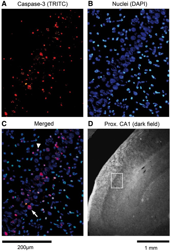
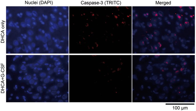
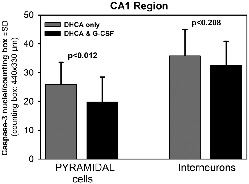
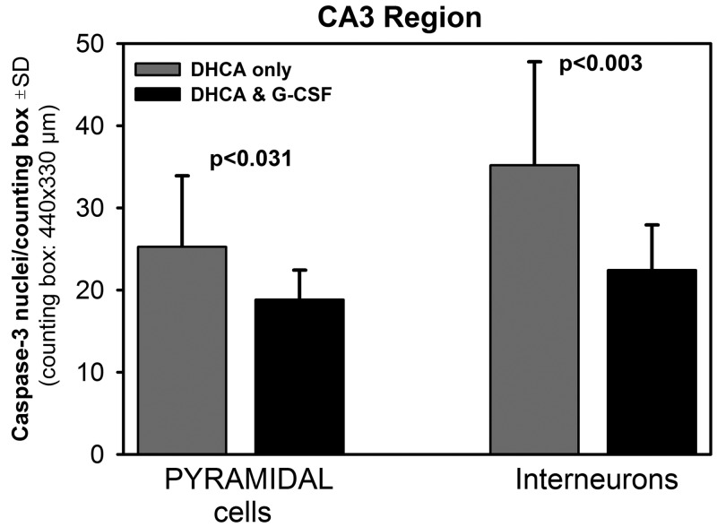
Methods: In a model of ischaemic brain injury, 4 male newborn piglets were anaesthetized and subjected to deep hypothermic circulatory arrest (DHCA) (cooled to 18°C, DHCA maintained for 60 min, rewarmed and recovered for 8-9 h), while 4 animals received G-CSF (34 µg/kg, intravenously) 2 h prior to the DHCA procedure. At the end of each experiment, the animals were perfused with a fixative, the hippocampus was extracted, cryoprotected, cut and the brain sections were immunoprocessed for activated caspase 3, a pro-apoptotic factor. Immunopositive neuronal nuclei were counted in multiple counting boxes (440 × 330 µm) centred on the CA1 or CA3 hippocampal regions and their mean numbers compared between the different treatment groups and regions.

Results: G-CSF pretreatment resulted in significantly lower counts of caspase 3-positive nuclei per counting box in both the CA1 [52.2 ± 9.3 (SD) vs 61.6 ± 8.4, P < 0.001] and CA3 (41.2 ± 6.9 vs 60.4 ± 16.4, P < 0.00002) regions of the hippocampus as compared to DHCA groups. The effects of G-CSF were significant for pyramidal cells of both regions and for interneurons in the CA3 region.

Conclusions: In an animal model of ischaemic brain injury, G-CSF reduces neuronal injury in the hippocampus, thus potentially having beneficial effect on neurologic outcomes.

Keywords: Apoptosis; Cardiac surgery; Caspase 3; Congenital heart disease; Developing brain; Hippocampus.


Figures

Figure 1
Figure 1
Caspase 3 immunoreactivity in the proximal CA1 region of the hippocampus of a piglet subjected to cardiopulmonary bypass with deep hypothermic cardiac arrest. (A) Image of caspase 3 immunostaining with tetramethyl rhodamine isothiocyanate (TRITC). (B) Same frame as in (A) but with blue nuclear staining (DAPI). (C) Merged frames shown in (A) and (B) demonstrate co-localization of caspase 3 in large (pyramidal) neurons and interneurons. Arrow indicates example of a caspase 3-positive pyramidal neuron; arrowhead indicates example of a caspase 3-positive interneuron. (D) Low-magnification, dark field image of a portion of the hippocampus with the white rectangle marking the location of the frame shown in panels (AC).
Figure 2
Figure 2
Comparison of caspase 3 immunoreactivity in the distal CA3 region of the hippocampus of a piglet subjected to cardiopulmonary bypass with deep hypothermic cardiac arrest (DHCA) (top) and a piglet pretreated with G-CSF prior to DHCA (bottom). The 3 panels for each case successively show blue nuclear DNA staining (DAPI), red caspase 3 immunostaining (TRITC), and a superimposition of both images (merged). In the animal pretreated with G-CSF (bottom), caspase 3 immunostaining is less extensive and occurs in fewer neurons than in the animal subjected to DHCA without pretreatment (top).
Figure 3
Figure 3
Mean counts of caspase 3-positive pyramidal cells and interneurons in the hippocampal CA1 region in the deep hypothermic circulatory arrest (DHCA)-only animals and the DHCA animals pretreated with G-CSF. There were 5.6 ± 2.5 (SD) cell counting boxes per animal and 4 animals per group. Significance levels were obtained using independent samples t-tests.
Figure 4
Figure 4
Mean counts of caspase 3-positive pyramidal cells and interneurons in the hippocampal CA3 region in the deep hypothermic circulatory arrest (DHCA)-only animals and the DHCA animals pretreated with G-CSF. There were 4.0 ± 1.3 (SD) cell counting boxes per animal and 4 animals per group. Significance levels were obtained using independent samples t-tests.

Similar articles

Cited by

References

    1. Arszovszki A, Borhegyi Z, Klausberger T.. Three axonal projection routes of individual pyramidal cells in the ventral CA1 hippocampus. Front Neuroanat 2014;8:53. - PMC - PubMed
    1. Graves AR, Moore SJ, Bloss EB, Mensh BD, Kath WL, Spruston N.. Hippocampal pyramidal neurons comprise two distinct cell types that are countermodulated by metabotropic receptors. Neuron 2012;76:776–89. - PMC - PubMed
    1. Barth AMI, Mody I.. Changes in hippocampal neuronal activity during and after unilateral selective hippocampal ischemia in vivo. J Neurosci 2011;31:851–60. - PMC - PubMed
    1. Schmidt-Kastner R, Freund TF.. Selective vulnerability of the hippocampus in brain ischemia. Neuroscience 1991;40:599–636. - PubMed
    1. Akai F, Yanagihara T.. Identity of the dorsal hippocampal region most vulnerable to cerebral ischemia. Brain Res 1993;603:87–95. - PubMed

MeSH terms

Substances