. 2017 Sep 29;8(1):202.
doi: 10.1186/s13287-017-0651-x.

Engineering human ventricular heart muscles based on a highly efficient system for purification of human pluripotent stem cell-derived ventricular cardiomyocytes

Affiliations

Engineering human ventricular heart muscles based on a highly efficient system for purification of human pluripotent stem cell-derived ventricular cardiomyocytes

Bin Li et al. Stem Cell Res Ther. .

Abstract

Background: Most infarctions occur in the left anterior descending coronary artery and cause myocardium damage of the left ventricle. Although current pluripotent stem cells (PSCs) and directed cardiac differentiation techniques are able to generate fetal-like human cardiomyocytes, isolation of pure ventricular cardiomyocytes has been challenging. For repairing ventricular damage, we aimed to establish a highly efficient purification system to obtain homogeneous ventricular cardiomyocytes and prepare engineered human ventricular heart muscles in a dish.

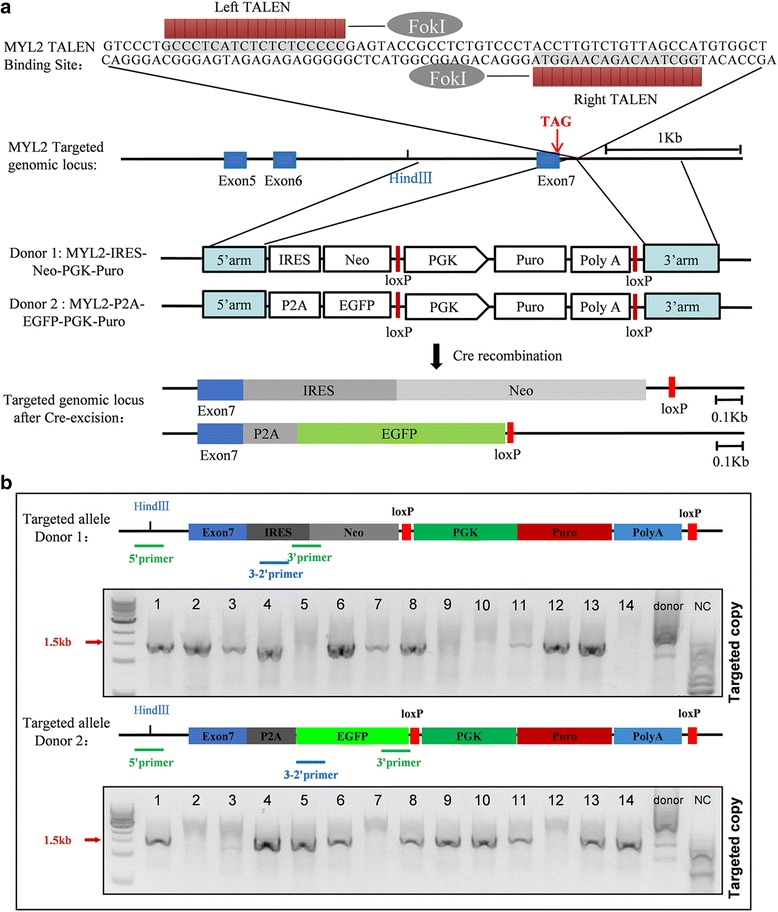
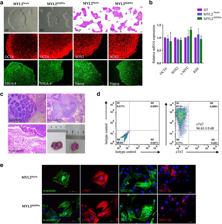
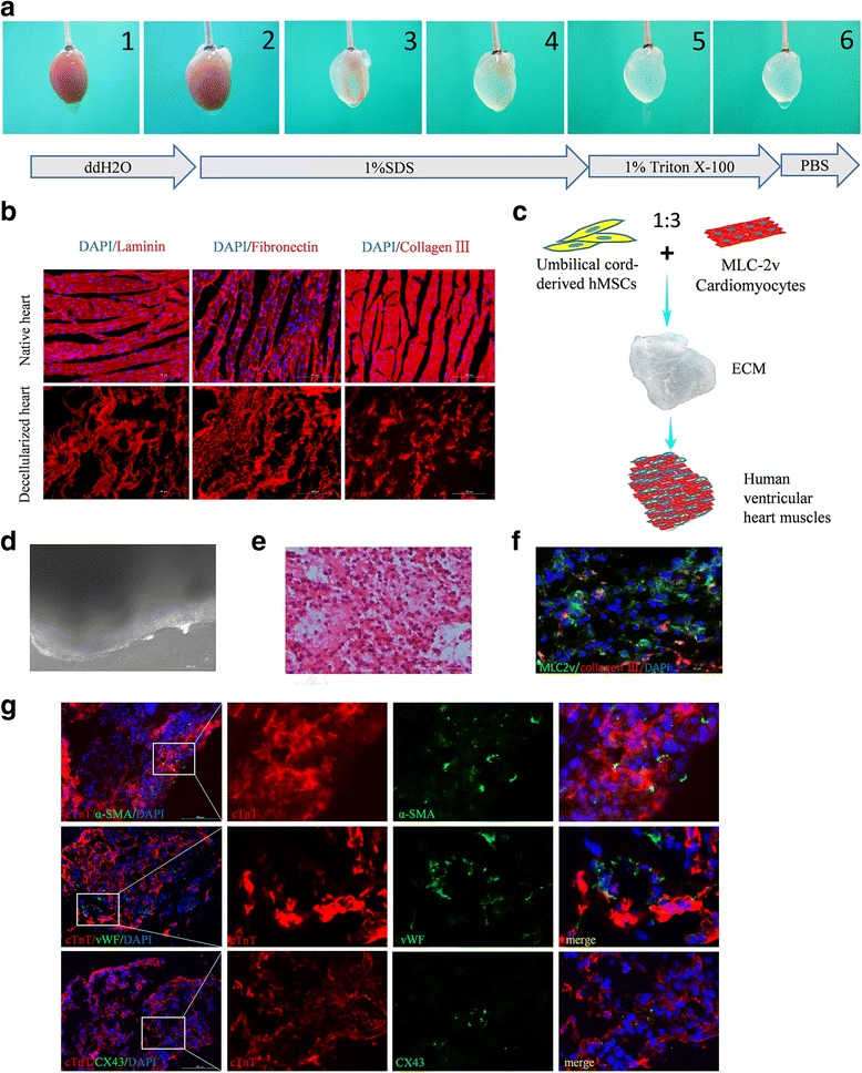
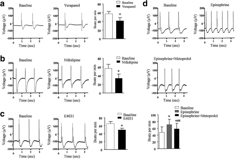
Methods: The purification system used TALEN-mediated genomic editing techniques to insert the neomycin or EGFP selection marker directly after the myosin light chain 2 (MYL2) locus in human pluripotent stem cells. Purified early ventricular cardiomyocytes were estimated by immunofluorescence, fluorescence-activated cell sorting, quantitative PCR, microelectrode array, and patch clamp. In subsequent experiments, the mixture of mature MYL2-positive ventricular cardiomyocytes and mesenchymal cells were cocultured with decellularized natural heart matrix. Histological and electrophysiology analyses of the formed tissues were performed 2 weeks later.

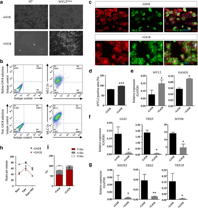
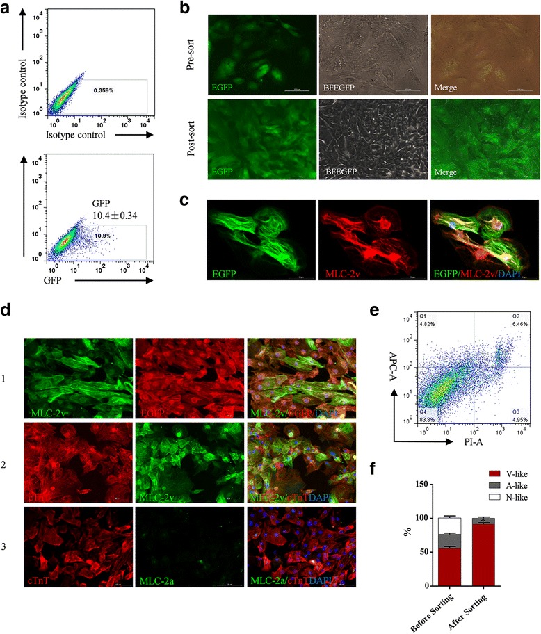
Results: Human ventricular cardiomyocytes were efficiently isolated based on the purification system using G418 or flow cytometry selection. When combined with the decellularized natural heart matrix as the scaffold, functional human ventricular heart muscles were prepared in a dish.

Conclusions: These engineered human ventricular muscles can be great tools for regenerative therapy of human ventricular damage as well as drug screening and ventricular-specific disease modeling in the future.

Keywords: Engineered human heart tissues; Engineered human ventricular heart muscles; Human pluripotent stem cells; Human ventricular cardiomyocytes; Myosin light chain 2; Myosin light chain 2v.


Conflict of interest statement

Ethics approval

All animal experiments in this study were approved by the Institutional Animal Care and Use Committee of the Fudan University (Committee approval number: 20140423) and were performed in accordance with the Guide for the Care and Use of Laboratory Animals (National Institutes of Health). All human stem cells research followed the ISSCR Guidelines for the Conduct of Human Embryonic Stem Cell Research. The human ESC line H7 used in this study was obtained from WiCell Research Institute under specific Material Transfer Agreement. The human iPSC line was derived from human skin fibroblasts with informed consent approved previously by the Bioethics Committee of Zhongshan Hospital affiliated to Fudan University.

Consent for publication

All authors have contributed to, read, and approved the final manuscript for submission.

Competing interests

The authors declare that they have no competing interests.

Publisher’s Note

Springer Nature remains neutral with regard to jurisdictional claims in published maps and institutional affiliations.

Figures

Fig. 1
Fig. 1
Schematics of the strategy for inserting the neomycin and EGFP selection cassette into the MYL2 locus. a TALENs targeting site for the MYL2 gene and the homologous recombination events. Red arrow indicates the stop codon of MYL2 gene. After introduction of a double-strand break near the MYL2 exon 7 after the stop codon by TALENs, an IRES-Neo (donor 1) or P2A-EGFP (donor 2) along with an excisable PGK-Puromycin drug selection cassette sequence was inserted into the MYL2 locus downstream of the TAG stop codon (middle panel). The bottom panel shows the targeted genomic locus after Cre-mediated excision of the Puro selection cassette. Blue box, exon of the MYL2 gene. b Nested-PCR strategy to identify successfully targeted clones. Both gels indicated 10 out of 14 clones had at least one copy of the targeted allele (targeting efficiency was 71.5%). MYL2 myosin light chain 2, TALEN transcription activator-like (TAL) effector nuclease, IRES internal ribosome entry site, P2A self-cleaving peptide sequence, Neo neomycin resistant cassette, EGFP enhanced green fluorescent protein, PGK phosphoglycerol kinase promoter, Puro puromycin resistance gene, polyA polyadenylation sequence, 5’arm 5’-homology arms, 3’arm 3’-homology arms
Fig. 2
Fig. 2
MYL2Neo/w and MYL2EGFP/w hESCs maintained pluripotency and the ability to differentiate into highly pure cardiomyocytes. a MYL2Neo/w and MYL2EGFP/w hESCs maintained pluripotent stem cell morphology (bright field (BF)), positive alkaline phosphatase (AP) staining, and expression of the pluripotency markers OCT4, SOX2, Nanog, and SSEA-4. Scale bars, 200 μm. b Quantitative PCR examining endogenous expression of the pluripotency factors relative to GAPDH in MYL2Neo/w and MYL2EGFP/w hESCs. Data obtained from three independent experiments. Wildtype hESCs (H7) used as controls. c Undifferentiated MYL2Neo/w and MYL2EGFP/w hESCs formed teratomas, which exhibited tissues of all three developmental germ layers: ectoderm (e.g., sebaceous gland cells), mesoderm (e.g., cartilage cells), and endoderm (e.g., gland cells). Scale bars, 200 μm. d Purity of MYL2Neo/w and MYL2EGFP/w hESC-derived cardiomyocytes detected by flow cytometry analysis of cardiac specific marker cTnT. e Immunostaining showing MYL2Neo/w and MYL2EGFP/w hESC-derived cardiomyocytes expressed cardiomyocyte specific proteins sarcomeric α-actinin, cTnT, MLC-2v, and MLC-2a. Scale bars, 50 μm. EGFP enhanced green fluorescent protein, MLC-2v cardiac ventricular isoform of myosin light chain-2, MYL2 myosin light chain 2
Fig. 3
Fig. 3
Effective enrichment of MLC-2v-positive human early ventricular cardiomyocytes based on the neomycin selection system. a Compared with the wildtype control (H7-derived cardiomyocytes), MYL2Neo/w hESC-derived cardiomyocytes show normal growth following G418 selection for 8–10 days after plating, while control cells died completely. b Flow cytometry analysis shows a higher percentage of MLC-2v-positive cells derived from MYL2Neo/w hESCs post G418 selection. c Representative immunofluorescence staining images reveal that MLC-2v was expressed in almost all of the G418 selected cTnT-positive MYL2Neo/w hESC-derived cardiomyocytes, while in control cells without G418 selection only partial cells showed MLC-2v expression. Scale bars, 100 μm. d Quantification of MLC-2v/cTnT expression ratio in MYL2Neo/w hESC-derived cardiomyocytes before and post G418 selection. n = 298, ***P < 0.001 by two-tailed Student’s t test. eg Quantitative PCR showing expression of ventricular markers MYL2 and HAND1 (e) was significantly upregulated, while atrial markers GJA5, TBX5, and MYH6 (f) and nodal markers SHOX2, TBX3, and TBX18 (g) were significantly reduced in MYL2Neo/w hESC-derived cardiomyocytes post G418 selection. Data shown as the mean ± SEM of three independent experiments. *P < 0.05; **P < 0.01; ns not statistically significant by two-tailed Student’s t test. h Quantification of beating rates for MYL2Neo/w hESC-derived cardiomyocytes in response to cardiac pharmaceutical reagents before and post G418 selection. i Percentages of ventricular-like, atrial-like, and nodal-like cells produced from MYL2Neo/w hESC-derived cardiomyocytes 30 days after cardiac differentiation before and post G418 selection as determined by single cell patch clamp. MYL2 myosin light chain 2, V-like ventricular-like cells, A-like atrial-like cells, N-like nodal-like cells
Fig. 4
Fig. 4
Effective enrichment of MLC-2v-positive human early ventricular cardiomyocytes based on the EGFP selection system. a FACS sorting showing positive cardiomyocytes derived from MYL2EGFP/w hESCs 25 days after cardiac differentiation. b Representative green fluorescence (EGFP), bright field (BFEGFP), and merged images of MYL2EGFP/w hESC-derived cardiomyocytes before and after FACS sorting. Scale bars, 100 μm. c Immunofluorescence microscopy showing coexpression of EGFP and MLC-2v in FACS sorted MYL2EGFP/w hESC-derived cardiomyocytes. Scale bars, 50 μm. d Double immunofluorescence staining showing coexpression of EGFP and MLC-2v in FACS sorted MYL2EGFP/w hESC-derived cardiomyocytes (1). MLC-2v was expressed in all of the cTnT-positive MYL2EGFP/w hESC-derived cardiomyocytes post FACS (2). MLC-2a only showed background expression level in the cTnT-positive MYL2EGFP/w hESC-derived cardiomyocytes post FACS (3). Scale bars, 100 μm. e Vitality assessment of the FACS selected MYL2EGFP/w-CMs by live cell staining for markers of apoptosis. Representative graph as detected by flow cytometry analysis. f Percentages of ventricular-like, atrial-like, and nodal-like cells produced from MYL2EGFP/w-CMs before and after FACS sorting as determined by single cell patch clamp. EGFP enhanced green fluorescent protein, MLC-2v cardiac ventricular isoform of myosin light chain-2, V-like ventricular-like cells, A-like atrial-like cells, N-like nodal-like cells
Fig. 5
Fig. 5
Coculture experiment showing that hPSC-derived ventricular cardiomyocytes electrically coupled with surrounding NRVMs. a Representative bright field phase contrast microscopic images (BF) and immunofluorescence staining of NRVM cultures (left panels) and NN-co-cultures, containing NRVMs and G418 selected MYL2Neo/w-CMs (right panels) in a 3:1 ratio. Cells were stained for cardiac troponin T (cTnT), human nuclear antigen (HNA), gap-junction protein connexin 43 (CX43), and DAPI. G418 selected MYL2Neo/w-CM nuclei were distinguished from rat nuclei by their positive staining for HNA. Scale bars, 50 μm. b Typical rhythmic spontaneous field potential recordings in NRVM cultures, NN-co-cultures, and NE-co-cultures (NRVMs and EGFP-positive MYL2EGFP/w-CMs). Quantification of (c) the beating frequency and (d) the percentage of irregular beats. NRVM neonatal rat ventricular myocyte, NN-co-culture MYL2Neo/w cardiomyocyte, NE-co-culture MYL2EGFP/w cardiomyocyte, ns not significant
Fig. 6
Fig. 6
Maturation medium leads to further maturation of the MLC-2v-positive ventricular-like cardiomyocytes. a Basal medium (control) and maturation medium (MM)-treated MYL2Neo/w-hiPSC-derived cardiomyocytes stained with α-actinin (green) and DAPI (blue). Scale bars, 50 μm. Right panels show enlarged views of the boxed areas in the left merged images, which show detailed α-actinin (green) staining patterns. Compared to MYL2Neo/w-hiPSC-derived cardiomyocytes cultured in basal medium (control), MM-treated MYL2Neo/w-hiPSC-derived cardiomyocytes show significant changes in cell size (b), sarcomere length (c), and number of multinuclear cells (d). n > 100 per condition. e Quantitative PCR showing higher transcriptional expression of the functional and mature genes in MM-treated MYL2Neo/w-CMs compared with control. Gene expressions shown normalized to GAPDH. Data shown as the mean ± SEM of three independent experiments. *P < 0.05; **P < 0.01; ***P <0.001 by two-tailed Student’s t test. f MYL2Neo/w-CMs show larger calcium transient amplitudes, faster upstroke, and faster decay velocities after culture with MM. n > 45 per condition
Fig. 7
Fig. 7
Generation and histological analysis of the human ventricular heart muscles. a Process of decellularization of rat hearts and preparation of natural heart ECM. b Major ECM compositions (laminin, fibronectin, and collagen III) of decellularized hearts and native rat hearts detected by immunofluorescence staining. c Schematic diagram of the preparation of engineered human heart muscles using MLC-2v-positive ventricular cardiomyocytes. d Representative image of the engineered human ventricular heart muscles in a light microscope. Scale bar, 200 μm. H&E staining (e) and coimmunostaining by anti-MLC-2v and anti-collagen III antibodies (f) of sections of constructed ventricular heart muscles. hPSC-derived ventricular cardiomyocytes are well distributed and attached on the native ECM. Scale bar, 100 μm. g Double immunofluorescence staining for cTnT/α-smooth muscle actin (α-SMA), cTnT/von Willebrand factor (vWF), and cTnT/CX43 in the engineered human ventricular heart muscles. Scale bars, 100 μm. ECM extracellular matrix, MLC-2v cardiac ventricular isoform of myosin light chain-2, hMSCs human mesenchymal stem cells
Fig. 8
Fig. 8
Electrophysiology analysis of human ventricular heart muscles. ad Ventricular heart muscles exhibited normal electrophysiology and responded to cardiac pharmaceutical reagents. MEA recording of the field potentials and contraction rate of the engineered human heart muscles before (baseline) and after treatment of verapamil (a), nifedipine (b), E4031 (c), and epinephrine and metoprolol (d). Data shown as the mean ± SEM of three independent experiments. *P < 0.05 compared with baseline; *##P < 0.05 compared with epinephrine alone by two-tailed Student’s t test

Similar articles

Cited by

References

    1. Lu ML, De Venecia T, Patnaik S, Figueredo VM. Atrial myocardial infarction: a tale of the forgotten chamber. Int J Cardiol. 2016;202:904–9. doi: 10.1016/j.ijcard.2015.10.070. - DOI - PubMed
    1. Laflamme MA, Murry CE. Heart regeneration. Nature. 2011;473:326–35. doi: 10.1038/nature10147. - DOI - PMC - PubMed
    1. Zwi L, Caspi O, Arbel G, Huber I, Gepstein A, Park IH, et al. Cardiomyocyte differentiation of human induced pluripotent stem cells. Circulation. 2009;120:1513–23. doi: 10.1161/CIRCULATIONAHA.109.868885. - DOI - PubMed
    1. Fluri DA, Tonge PD, Song H, Baptista RP, Shakiba N, Shukla S, et al. Derivation, expansion and differentiation of induced pluripotent stem cells in continuous suspension cultures. Nat Methods. 2012;9:509–16. doi: 10.1038/nmeth.1939. - DOI - PMC - PubMed
    1. Burridge PW, Matsa E, Shukla P, Lin ZC, Churko JM, Ebert AD, et al. Chemically defined generation of human cardiomyocytes. Nat Methods. 2014;11:855–60. doi: 10.1038/nmeth.2999. - DOI - PMC - PubMed

Publication types

MeSH terms