. 2017 Sep 29;8(1):737.
doi: 10.1038/s41467-017-00917-6.

Boosting the down-shifting luminescence of rare-earth nanocrystals for biological imaging beyond 1500 nm

Affiliations

Boosting the down-shifting luminescence of rare-earth nanocrystals for biological imaging beyond 1500 nm

Yeteng Zhong et al. Nat Commun. .

Abstract

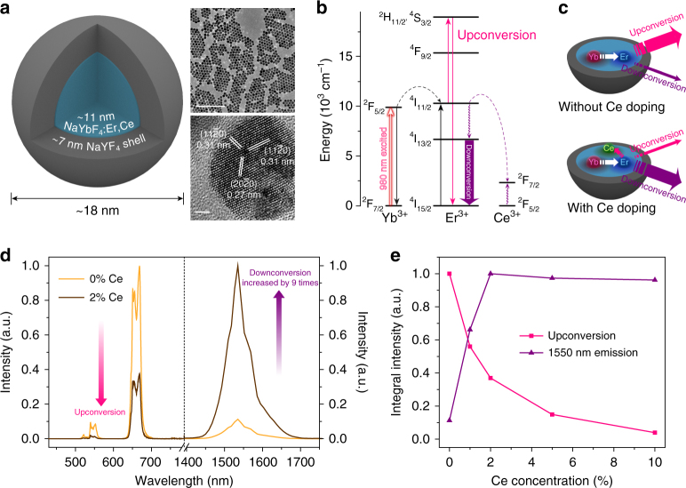
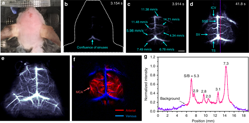
In vivo fluorescence imaging in the near-infrared region between 1500-1700 nm (NIR-IIb window) affords high spatial resolution, deep-tissue penetration, and diminished auto-fluorescence due to the suppressed scattering of long-wavelength photons and large fluorophore Stokes shifts. However, very few NIR-IIb fluorescent probes exist currently. Here, we report the synthesis of a down-conversion luminescent rare-earth nanocrystal with cerium doping (Er/Ce co-doped NaYbF4 nanocrystal core with an inert NaYF4 shell). Ce doping is found to suppress the up-conversion pathway while boosting down-conversion by ~9-fold to produce bright 1550 nm luminescence under 980 nm excitation. Optimization of the inert shell coating surrounding the core and hydrophilic surface functionalization minimize the luminescence quenching effect by water. The resulting biocompatible, bright 1550 nm emitting nanoparticles enable fast in vivo imaging of blood vasculature in the mouse brain and hindlimb in the NIR-IIb window with short exposure time of 20 ms for rare-earth based probes.Fluorescence imaging in the near-infrared window between 1500-1700 nm (NIR-IIb window) offers superior spatial resolution and tissue penetration depth, but few NIR-IIb probes exist. Here, the authors synthesize rare earth down-converting nanocrystals as promising fluorescent probes for in vivo imaging in this spectral region.


Conflict of interest statement

The authors declare no competing financial interests.

Figures

Fig. 1
Fig. 1
Ce3+ doped rare-earth nanoparticles with enhanced NIR-IIb luminescence. a Schematic design of a NaYbF4:Er,Ce@NaYF4 core-shell nanoparticle (left) with corresponding large scale TEM image (upper right, scale bar = 200 nm) and HRTEM image (lower right, scale bar = 2 nm). b Simplified energy-level diagrams depicting the energy transfer between Yb3+, Er3+, and Ce3+ ions. c Schematic illustration of the proposed energy-transfer mechanisms in Er-RENPs with and without Ce3+ doping. d Upconversion and downconversion luminescence spectra of the Er-RENPs with 0 and 2% Ce3+ doping. e Schematic representation of Ce3+ doping concentration and corresponding upconversion and downconversion emission intensity of the Er-RENPs upon 980 nm excitation
Fig. 2
Fig. 2
Surface modification of the Er-RENPs. a Schematic illustration outlining the PMH coating and PEGylation procedure for the Er-RENPs (Er-RENPs@PMH-PEG). b DLS spectra of PMH capped RENPs before and after PEGylation. c DLS spectra demonstrating the well dispersibility of RENPs@PMH-PEG in different concentration of PBS solution. d Downconversion emission intensity of Er-RENPs@PMH-PEG in 1x PBS and 37 °C FBS solution as a function of days. The inset showed 1550 nm luminescence images of Er-RENPs@PMH-PEG in 1x PBS at 0th and 7th day. e Downconversion luminescence spectra of oleic acid-capped Er-RENPs dispersed in cyclohexane and Er-RENPs@PMH-PEG dispersed in water
Fig. 3
Fig. 3
Reducing aqueous quenching effect by controlling the inert shell thickness of Er-RENPs. a Schematic illustration of the proposed quenching mechanisms of Er-RENPs in aqueous solution. b Schematic representation of shell thickness and corresponding 1550 nm downconversion emission intensity of the Er-RENPs in organic and aqueous phase upon 980 nm excitation. c Quenching rate of upconversion and downconversion emission as a function of shell thickness (from 3.2 nm to 8.1 nm). Three surface coating experiments were performed for each Er-RENPs sample. All data are presented as means ± s.d
Fig. 4
Fig. 4
Fast in vivo brain imaging with Er-RENPs@PMH-PEG in the NIR-IIb region. a Color photograph of a C57Bl/6 mouse (with hair shaved off) preceding NIR-IIb fluorescence imaging. bd Time-course NIR-IIb brain fluorescence images (exposure time: 20 ms) showing the perfusion of RENPs into various cerebral vessels. The blood-flow velocities of cerebral vessels are given in c (scale bar corresponds to bd: 2 mm). e, f Cerebral vascular image (exposure time: 20 ms) in NIR-IIb region with corresponding PCA overlaid image f showing arterial (red) and venous (blue) vessels. g SBR analysis of NIR-IIb cerebrovascular image d by plotting the cross-sectional intensity profiles

Similar articles

Cited by

References

    1. Choi HS, et al. Targeted zwitterionic near-infrared fluorophores for improved optical imaging. Nat. Biotechnol. 2013;31:148–153. doi: 10.1038/nbt.2468. - DOI - PMC - PubMed
    1. Hong G, et al. Ultrafast fluorescence imaging in vivo with conjugated polymer fluorophores in the second near-infrared window. Nat. Commun. 2014;5:4206. - PubMed
    1. Zhu S, et al. Molecular imaging of biological systems with a clickable dye in the broad 800- to 1,700-nm near-infrared window. Proc. Natl Acad. Sci. USA. 2017;114:962–967. doi: 10.1073/pnas.1617990114. - DOI - PMC - PubMed
    1. Hong G, et al. Multifunctional in vivo vascular imaging using near-infrared II fluorescence. Nat. Med. 2012;18:1841–1846. doi: 10.1038/nm.2995. - DOI - PMC - PubMed
    1. Welsher K, Sherlock SP, Dai H. Deep-tissue anatomical imaging of mice using carbon nanotube fluorophores in the second near-infrared window. Proc. Natl Acad. Sci. USA. 2011;108:8943–8948. doi: 10.1073/pnas.1014501108. - DOI - PMC - PubMed

Publication types