. 2017 Sep 29;8(1):748.
doi: 10.1038/s41467-017-00751-w.

Foxp3+ Tregs are recruited to the retina to repair pathological angiogenesis

Affiliations

Foxp3+ Tregs are recruited to the retina to repair pathological angiogenesis

Devy Deliyanti et al. Nat Commun. .

Abstract

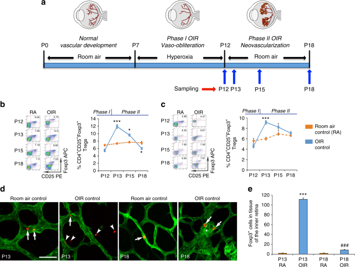
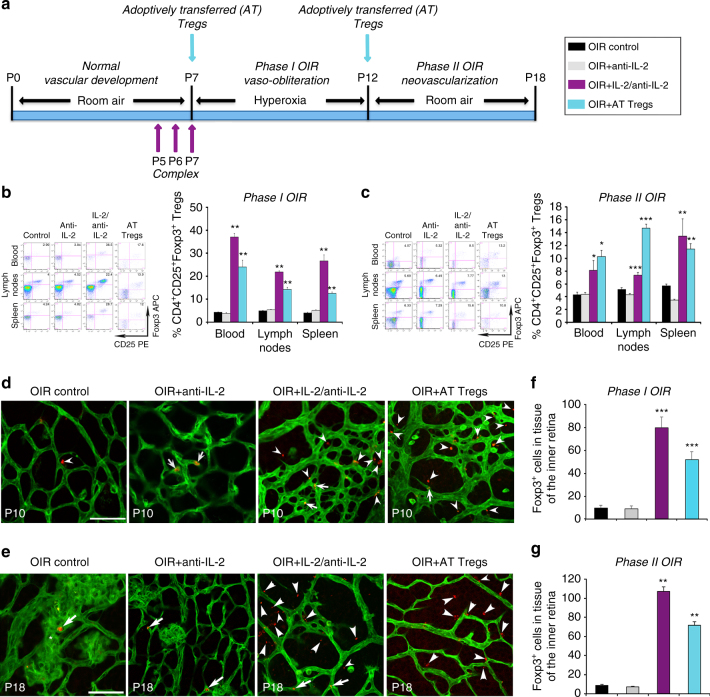
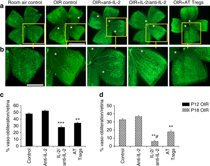
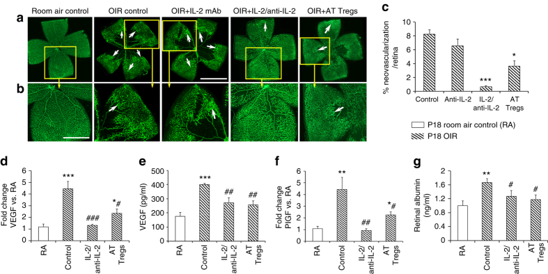
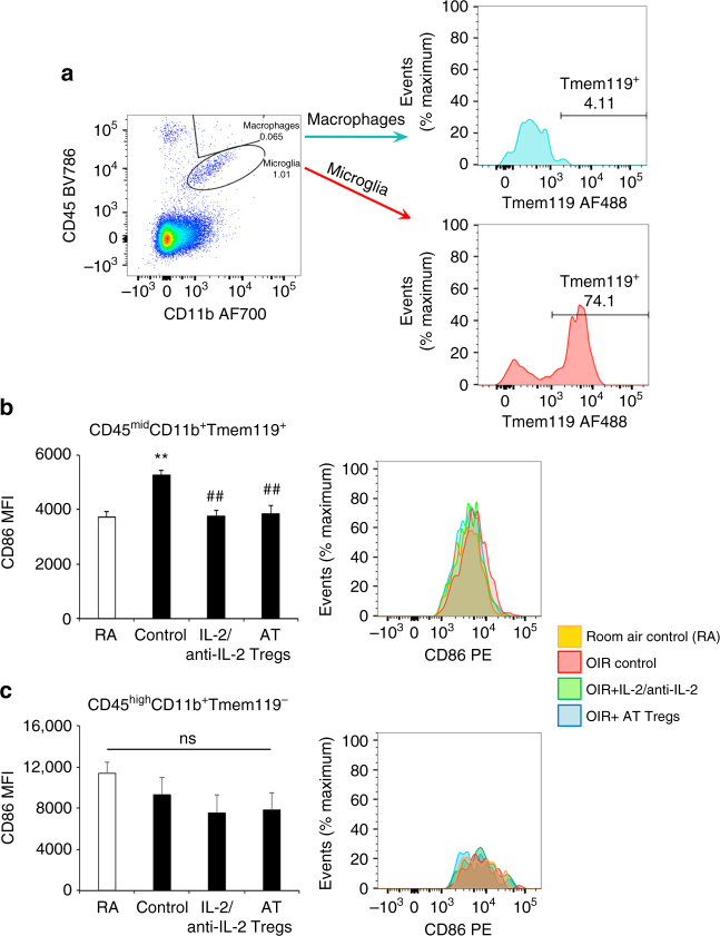
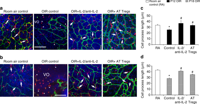
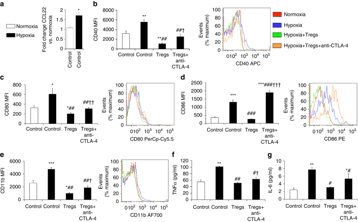
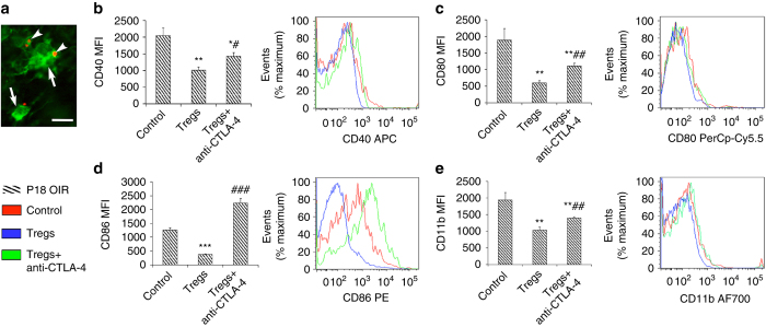
Neovascular retinopathies are major causes of vision loss; yet treatments to prevent the condition are inadequate. The role of regulatory T cells in neovascular retinopathy is unknown. Here we show that in retinopathy regulatory T cells are transiently increased in lymphoid organs and the retina, but decline when neovascularization is established. The decline is prevented following regulatory T cells expansion with an IL-2/anti-IL-2 mAb complex or the adoptive transfer of regulatory T cells. Further, both approaches reduce vasculopathy (vaso-obliteration, neovascularization, vascular leakage) and alter the activation of Tmem119+ retinal microglia. Our in vitro studies complement these findings, showing that retinal microglia co-cultured with regulatory T cells exhibit a reduction in co-stimulatory molecules and pro-inflammatory mediators that is attenuated by CTLA-4 blockade. Collectively, we demonstrate that regulatory T cells are recruited to the retina and, when expanded in number, repair the vasculature. Manipulation of regulatory T cell numbers is a previously unrecognized, and promising avenue for therapies to prevent blinding neovascular retinopathies.The local immune responses in the eye are attenuated to preserve sight. Surprisingly, Deliyanti et al. show that regulatory T cells (Tregs) take an active role in protecting the eye from neovascularization in oxygen-induced retinopathy, and that interventions that augment the retinal Treg numbers reduce neovascular retinopathy in mice.


Conflict of interest statement

The authors declare no competing financial interests.

Figures

Fig. 1
Fig. 1
Tregs are transiently increased in OIR and penetrate the retina. a Cohorts of C57BL/6J mice were studied during phase I OIR at postnatal day (P) 12 and phase II OIR at P13, P15, and P18. Comparisons were made to age-matched room air controls (RA). FACS of pooled lymph nodes b and spleen c revealed that Foxp3+ Tregs were increased in phase II OIR at P13 and declined to the level of RA or below by P18 (enlarged FACS plots in Supplementary Fig. 2). Fluorochromes are phycoerythrin (PE) and allophycocyanin (APC). n = 13 RA mice at P13 and P18. n = 7 OIR mice at P13 and P18. *P < 0.05 and ***P < 0.001 to age-matched RA (one-way ANOVA with Mann–Whitney U test). d Representative confocal images of flat mounted retina from Foxp3rfp mice at P13 and P18 labeled with isolectin (green) to show blood vessels. Scale bar, 50 μm. In RA, Foxp3+ cells (red) were mainly located within blood vessels (arrows). In OIR at P13, Foxp3+ cells were increased and mainly located in retinal tissue (arrowheads). In OIR at P18, the number of Foxp3+ cells had declined and were localized to tufts of neovascular blood vessels (asterisk) with few cells in retinal tissue. e Foxp3+ cells in tissue of the inner retina (one-way ANOVA with Mann–Whitney U test). ***P < 0.001 to all groups. ### P < 0.001 to RA. n = 16 RA mice and OIR mice at P13. n = 16 RA mice and 12 OIR mice at P18. Values are expressed as mean ± s.e.m
Fig. 2
Fig. 2
Expanding Treg numbers increased their abundance in OIR and retina. a C57BL/6J mice were administered an IL-2/anti-IL-2 mAb complex at postnatal day (P) 5, P6, and P7. Separate cohorts of OIR mice received purified splenic Tregs by adoptive transfer (AT) at P7 and P12. Mice were studied in phase I OIR and phase II OIR and comparisons made to age-matched OIR control mice or OIR mice administered an anti-IL-2 control mAb. FACS revealed that in phase I OIR at P12 b and phase II OIR at P18 c the numbers of Tregs in blood, pooled lymph nodes, and spleen were increased with both treatments (enlarged FACS plots in Supplementary Fig. 3). Fluorochromes are phycoerythrin PE) and allophycocyanin (APC). *P < 0.05, **P < 0.01, and ***P < 0.001 to OIR controls and OIR + anti-IL-2 mAb controls (one-way ANOVA with Mann–Whitney U test). n = 6 mice/OIR control and OIR + anti-IL-2 group and 7 mice/OIR-treated group at P12 and P18. Representative confocal images of flat mounted retinas from Foxp3rfp mice labeled with isolectin (green) to show blood vessels. In phase I OIR at P10 d and phase II OIR at P18 e, few Foxp3+ cells (red) were present in retinal tissue and (arrowhead) and blood vessels (arrows). Asterisk denotes neovascular tuft. In OIR-treated groups, Foxp3+ cells were increased in retinal tissue (arrowheads). Scale bar, 50 μm. f, g Foxp3+ cells in tissue of the inner retina (one-way ANOVA with Mann–Whitney U test). **P < 0.01 and ***P < 0.001 to OIR control groups. n = 13 mice/OIR control group and 9 mice/OIR-treated group at P10. n = 12 OIR mice, 9 OIR + anti-IL-2 mice and 7 mice/OIR-treated groups at P18. Values are expressed as mean ± s.e.m
Fig. 3
Fig. 3
Expanding Treg numbers reduced retinal vaso-obliteration in OIR. a Representative confocal images of flat mounted retina from C57BL/6J mice at postnatal day (P) 12 in phase I OIR and labeled with isolectin (green) to show blood vessels. Scale bar, 0.5 mm. b One quadrant (yellow box) from each retina has been enlarged. Scale bar, 0.25 mm. Retinas from room air control mice show normal vascularization, while retinas from OIR controls and OIR + anti-IL-2 mAb have vaso-obliterated blood vessels (asterisks) in the central retina. Mice treated with an IL-2/anti-IL-2 mAb complex or adoptively transferred (AT) Tregs have reduced retinal vaso-obliteration. Quantitation in phase I OIR at P12 c and phase II OIR at P18 d **P < 0.01 and ***P < 0.001 to OIR control groups. # P < 0.05 to OIR + AT Tregs (one-way ANOVA with Mann–Whitney U test). n = 9 mice/group with three litters examined. Values are mean ± s.e.m
Fig. 4
Fig. 4
Expanding Treg numbers reduced retinal neovascularization in OIR. a Representative confocal images of flat mounted retina from C57BL/6J mice at postnatal day (P) 18 in phase II OIR and labeled with isolectin to show blood vessels (green). Scale bar, 0.5 mm. b One quadrant (yellow box) from each retina has been enlarged. Scale bar, 0.25 mm. Retinas from room air control (RA) mice exhibit normal vascularization, while retinas from OIR controls and OIR + anti-IL-2 mAb display marked neovascularization (arrows). OIR mice treated with an IL-2/anti-IL-2 mAb complex or adoptively transferred (AT) Tregs have reduced retinal neovascularization. c Neovascularization is expressed as a percentage of total retinal area. *P < 0.05, ***P < 0.001 to OIR control groups (one-way ANOVA with Mann–Whitney U test). n = 7 room air control mice and 9 mice/OIR group with three litters examined. d vegfa mRNA, e VEGF protein, f placental growth factor mRNA, and g vascular leakage were increased in retina from OIR mice compared to RA, and in OIR were reduced by both treatments. *P < 0.05, **P < 0.01, and ***P < 0.001 to RA. # P < 0.05, ## P < 0.01, and ### P < 0.001 to OIR control (one-way ANOVA with Mann–Whitney U test). n = 5 room air control mice and 7 mice/OIR group. Values are expressed as mean ± s.e.m
Fig. 5
Fig. 5
Expanding Treg numbers altered the activation state of microglia. Flow cytometry of retina from C57BL/6J mice at postnatal day 10 in phase I OIR. Fluorochromes are AF700, AF488 and phycoerythrin (PE). a FACS plot schema showing cell populations in OIR retina revealed by CD45 and CD11b staining. Microglia (CD45midCD11b+) rather than macrophages (CD45highCD11b+) predominate and Tmem119 is restricted to CD45midCD11b+ microglia. b Mean fluorescence intensity (MFI) of CD86 in CD45midCD11b+Tmem119+ microglia showing an increase with OIR compared to room air controls (RA), and a reduction in OIR mice following treatment with the IL-2/anti-IL-2 mAb complex and adoptively transferred (AT) Tregs. c MFI of CD86 in CD45highCD11b+Tmem119 macrophages showing no change with OIR compared to RA or the treatments. **P < 0.01 to RA. ## P < 0.01 to OIR control (one-way ANOVA with Mann–Whitney U test). NS, not significant. n = 5 mice group. Values are expressed as mean ± s.e.m
Fig. 6
Fig. 6
Expanding Treg numbers preserved the ramified phenotype of microglia. Representative confocal images of flat mounted retina from C57BL/6J mice showing blood vessels (isolectin, green), microglia (Iba1+, red), and nuclei (DAPI, blue). Scale bar, 100 μm. In room air controls (RA) at a P12 and b P18, Iba1+ cells with long ramified processes (arrows) predominate, while in OIR at both time points, most Iba1+ cells exhibit short cell processes (asterisks) and are found in the vaso-obliterated (VO) retina. In OIR mice treated with the IL-2/anti-IL-2 mAb complex or the adoptive transfer (AT) of Tregs, the number of Iba1+ cells with long ramified cell processes (arrows) predominate. The primary cell process of each Iba1+ cell per field of retina was quantitated at c P12 and d P18. *P < 0.05 to RA. # P < 0.05 to OIR control (one-way ANOVA with Mann–Whitney U test). n = 6 room air control mice and 8 mice/OIR group. Values are expressed as mean ± s.e.m
Fig. 7
Fig. 7
In vitro Tregs alter the activation of retinal microglia. a Primary cultures of microglia were established from the retinas of 10 to 12-day-old C57BL/6J mice. Quantitative real-time PCR revealed that ccl22 mRNA levels were increased by hypoxia (0.5% O2). *P < 0.05. bg Retinal microglia were exposed to hypoxia (0.5% O2) and co-cultured with Tregs and a blocking anti-CTLA-4 mAb (10 μg/ml). Flow cytometry was performed and data are expressed as mean fluorescence intensity (MFI) for b CD40, c CD80, d CD86, and e CD11b. Fluorochromes are allophycocyanin (APC), PerCp-Cy5.5, phycoerythrin (PE), and AF700. *P < 0.05, **P < 0.01, and ***P < 0.001 to normoxia control. ## P < 0.01 and ### P < 0.001 to hypoxia control. P < 0.05, †† P < 0.01, and ††† P < 0.001 to hypoxia + Tregs. In the cell supernatant, an ELISA was used to measure f TNFα and g IL-6 levels in cell supernatant. *P < 0.05 and **P < 0.01 to normoxia control. # P < 0.05 and ## P < 0.01 to hypoxia control. P < 0.05 to hypoxia + Tregs. Data are representative of two independent experiments each containing three replicates (one-way ANOVA with Student’s t test). Values are expressed as mean ± s.e.m
Fig. 8
Fig. 8
The activation state of OIR microglia is altered by Tregs. a Representative confocal image of a flat mounted retina from Foxp3rfp mice at postnatal day (P) 18 with OIR and treated with the IL-2/anti-Il-2 mAb complex. Foxp3+ cells (red, arrowheads) contact microglia (Iba1 immunolabeling in green, arrows). Scale bar, 40 μm. be Microglia from retinas of OIR mice at P18 were co-cultured with Tregs purified from adult mouse spleen and incubated with a blocking anti-CTLA-4 mAb (10 μg/ml). Flow cytometry was performed and mean fluorescence intensity (MFI) quantitated for b CD40, c CD80, d CD86, and e CD11b. Fluorochromes are allophycocyanin (APC), PerCp-Cy5.5, phycoerythrin (PE), and AF700. *P < 0.05, **P < 0.01, and **P < 0.001 to OIR control. # P < 0.05, ## P < 0.01, and ### P < 0.001 to OIR + Tregs. Data are representative of two independent experiments each containing three replicates (one-way ANOVA with Student’s t test). Values are expressed as mean ± s.e.m

Comment in

References

    1. Simo R, Hernandez C. Novel approaches for treating diabetic retinopathy based on recent pathogenic evidence. Prog. Retin. Eye Res. 2015;48:160–180. doi: 10.1016/j.preteyeres.2015.04.003. - DOI - PubMed
    1. Sapieha P, et al. Retinopathy of prematurity: understanding ischemic retinal vasculopathies at an extreme of life. J. Clin. Invest. 2010;120:3022–3032. doi: 10.1172/JCI42142. - DOI - PMC - PubMed
    1. Agarwal A, et al. Management of neovascular age-related macular degeneration: current state-of-the-art care for optimizing visual outcomes and therapies in development. Clin. Ophthalmol. 2015;9:1001–1015. - PMC - PubMed
    1. Hellstrom A, Smith LE, Dammann O. Retinopathy of prematurity. Lancet. 2013;382:1445–1457. doi: 10.1016/S0140-6736(13)60178-6. - DOI - PMC - PubMed
    1. Campbell K. Intensive oxygen therapy as a possible cause of retrolental fibroplasia: a clinical approach. Med. J. Aust. 1951;2:48–50. - PubMed

Publication types