. 2017 Sep 29;7(1):12454.
doi: 10.1038/s41598-017-12378-4.

Label-free detection of cellular drug responses by high-throughput bright-field imaging and machine learning

Affiliations

Label-free detection of cellular drug responses by high-throughput bright-field imaging and machine learning

Hirofumi Kobayashi et al. Sci Rep. .

Abstract

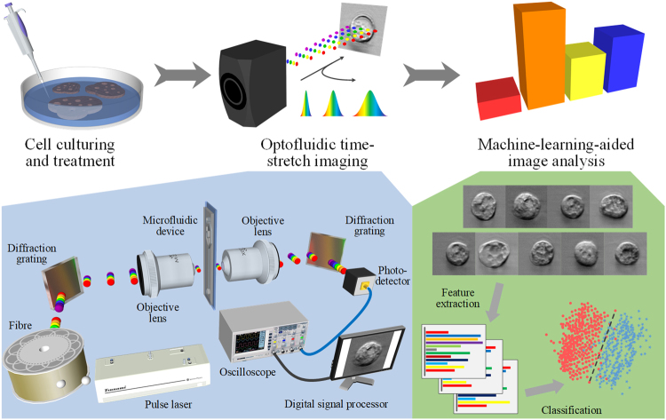
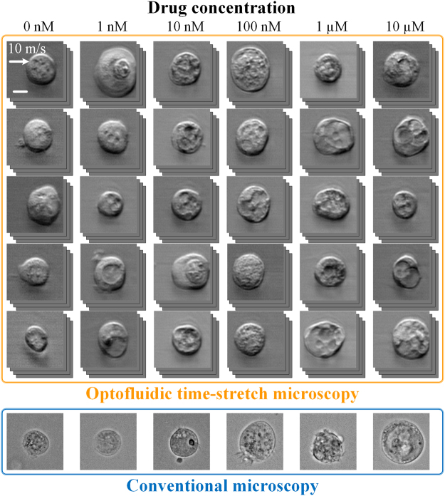
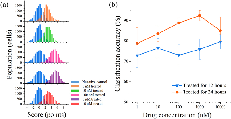
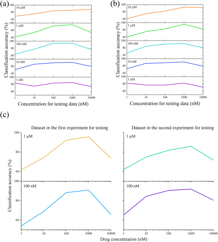
In the last decade, high-content screening based on multivariate single-cell imaging has been proven effective in drug discovery to evaluate drug-induced phenotypic variations. Unfortunately, this method inherently requires fluorescent labeling which has several drawbacks. Here we present a label-free method for evaluating cellular drug responses only by high-throughput bright-field imaging with the aid of machine learning algorithms. Specifically, we performed high-throughput bright-field imaging of numerous drug-treated and -untreated cells (N = ~240,000) by optofluidic time-stretch microscopy with high throughput up to 10,000 cells/s and applied machine learning to the cell images to identify their morphological variations which are too subtle for human eyes to detect. Consequently, we achieved a high accuracy of 92% in distinguishing drug-treated and -untreated cells without the need for labeling. Furthermore, we also demonstrated that dose-dependent, drug-induced morphological change from different experiments can be inferred from the classification accuracy of a single classification model. Our work lays the groundwork for label-free drug screening in pharmaceutical science and industry.


Conflict of interest statement

The authors declare that they have no competing interests.

Figures

Figure 1
Figure 1
Workflow of the label-free detection of drug-induced morphological variations in cancer cells with optofluidic time-stretch microscopy. The insets show the schematics of optofluidic time-stretch microscopy and machine-learning-aided image analysis. As schematically shown in the figure, the procedure of our method can be divided into three parts: (i) cell culturing and treatment, (ii) optofluidic time-stretch imaging, and (iii) machine-learning-aided image analysis. In the first part, cells of interest are cultured and treated by a drug. In the second part, the treated cells are subject to high-throughput bright-field imaging. In the last part, machine learning algorithm is applied to the images for the identification of their morphological variations induced by drug treatment. While the morphological change in a single cell is miniscule, the large number of bright-field single-cell images can render the morphological change discovered by machine learning statistically significant and robust.
Figure 2
Figure 2
Image libraries of drug-treated and -untreated MCF-7 cells under our optofluidic time-stretch microscope (flowing at a speed of 10 m/s). Compared with the static images obtained by a conventional microscope, despite the high flow speed, the optofluidic time-stretch microscope can acquire blur-free images of cells with decent image quality. Scale bars: 10 µm.
Figure 3
Figure 3
Classification of drug-treated and -untreated cancer cells. (a) Histograms of SVM classification scores for MCF-7 cells treated with various concentrations of paclitaxel for 24 hours. Each population consists of up to 10,000 cells. (b) Classification accuracy at various drug concentrations and incubation times. The error bars represent standard errors of the cross-validation estimation of average classification accuracy (n = 4).
Figure 4
Figure 4
Calculating maximum mean discrepancy (MMD) between the negative control and drug-treated cell population. (a) Illustration of the maximum mean discrepancy (MMD). (b) MMD between the negative control and drug-treated cell population at each drug concentration. Trial 1: data from the first experiment. Trial 2: data from the second experiment. (c) MMD of each feature in trial 1 at 1 µM and trial 2 at 100 nM. At these concentrations, the MMD in the whole feature space is the largest in each experiment. Features with a higher score of the MMD in both trials are highly correlated, indicating that the significant features were consistent in both experiments. The color scale represents feature index, showing types of morphological changes that undergo larger scores of the MMD. (d) Classification accuracy with a reduced number of features. Lower MMD features were removed based on the ranking of the MMD for each feature in the classification between the negative control and the dataset at 1 µM in the first experiment (top). The classification accuracy was maintained over 90% with more than 100 features (bottom). The color scale is consistent with that in Fig. 4c. Feature ranking and the number of remaining features are illustrated in logarithmic scale. The error bars represent standard errors of the cross-validation estimation of average classification accuracy (n = 10).
Figure 5
Figure 5
Classification accuracy using single SVM models. Classification accuracy produced by the SVM models in the first experiment (a) and the second experiment (b). Each row demonstrates the classification accuracy at each drug concentration produced by a single SVM model. (c) Evaluation of single SVM models across different experiments. Each row demonstrates the classification accuracy at each drug concentration produced by a single SVM model trained with the data from 1 µM in the first experiment (upper) and 100 nM in the second experiment (lower). Each column demonstrates the testing data from different trial of experiments.

References

    1. Usaj MM, et al. High-content screening for quantitative cell biology. Trends Cell Biol. 2016;26:598–611. doi: 10.1016/j.tcb.2016.03.008. - DOI - PubMed
    1. Heynen-Genel S, Pache L, Chanda SK, Rosen J. Functional genomic and high-content screening for target discovery and deconvolution. Expert Opin. Drug Discov. 2012;7:955–968. doi: 10.1517/17460441.2012.711311. - DOI - PMC - PubMed
    1. Futamura Y, et al. Morphobase, an encyclopedic cell morphology database, and its use for drug target identification. Chem. Biol. 2012;19:1620–1630. doi: 10.1016/j.chembiol.2012.10.014. - DOI - PubMed
    1. Loo LH, Wu LF, Altschuler SJ. Image-based multivariate profiling of drug responses from single cells. Nat. Methods. 2007;4:445–453. - PubMed
    1. Perlman, Z. E. et al. Multidimensional drug profiling by automated microscopy. Science306; (2004). - PubMed

Publication types

MeSH terms

Substances