. 2017 Sep 29;7(1):12426.
doi: 10.1038/s41598-017-10962-2.

Human Amniotic Membrane Mesenchymal Stem Cells inhibit Neutrophil Extracellular Traps through TSG-6

Affiliations

Human Amniotic Membrane Mesenchymal Stem Cells inhibit Neutrophil Extracellular Traps through TSG-6

Fátima Sofía Magaña-Guerrero et al. Sci Rep. .

Abstract

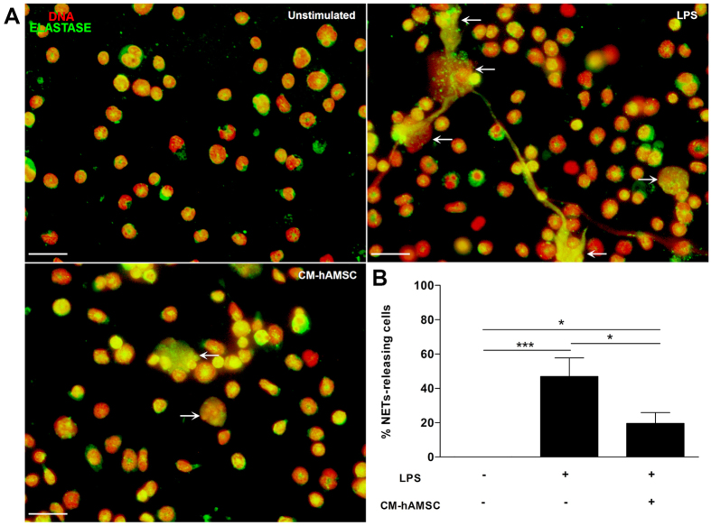
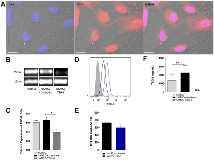
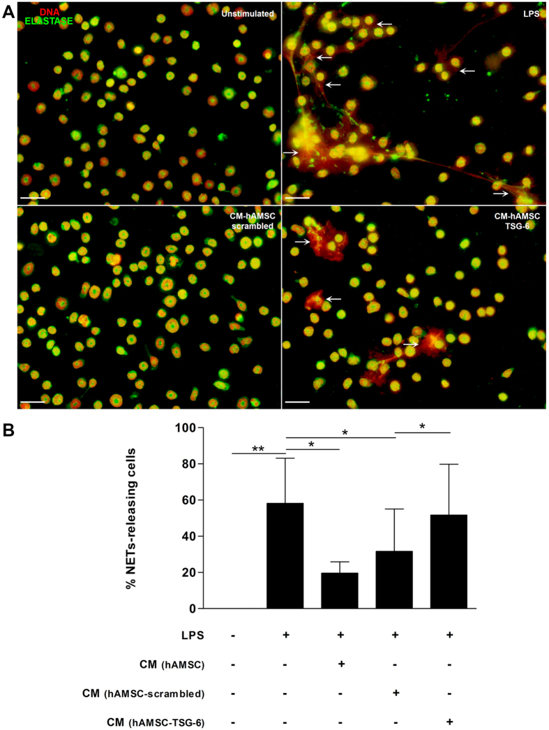
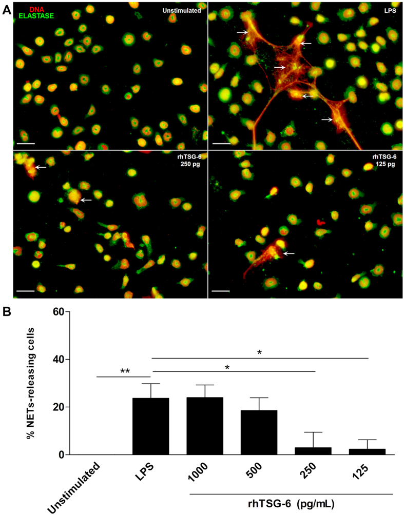
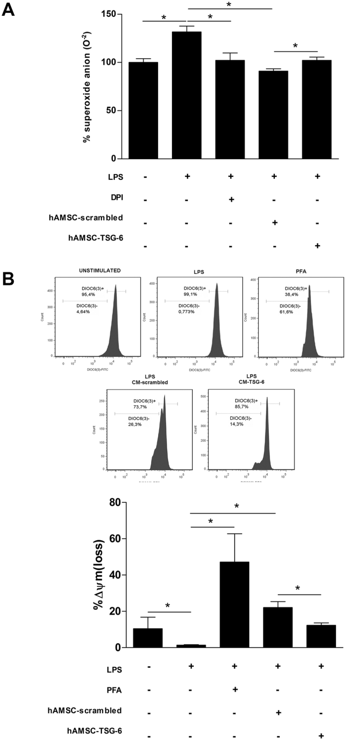
The mesenchymal stem cells obtained from human amniotic membrane (hAMSC) possess immunosuppressive functions through soluble factors such as prostanoids and proteins; thus, they have been proposed to ameliorate inflammatory processes. On the other hand, activated neutrophils are cells of the first line of immune defense that are able to release extracellular traps (NETs). NETs are formed of DNA and granular components; however, the excessive release of NETs is associated with the development of autoimmune and chronic inflammatory diseases. In this study, we identified that conditioned medium (CM) from hAMSC was able to diminish NETs release, as well as the production of reactive oxygen species (ROS) and the mitochondrial membrane potential from LPS-stimulated mouse bone marrow-derived neutrophils (BMN). Interestingly, NETs inhibition, ROS levels decrease and mitochondrial membrane potential loss were reverted when LPS-stimulated murine derived BMN were exposed to the CM from hAMSC transfected with TSG-6-siRNA. Finally, rhTSG6 was able to significantly diminish NETs release in BMN. These data suggest an inhibition mechanism of NETs ROS-dependent in which TSG-6 participates. Consequently, we propose the hAMSC use as a therapeutic candidate in the treatment of inflammatory diseases in which NETs are involved.


Conflict of interest statement

The authors declare that they have no competing interests.

Figures

Figure 1
Figure 1
Cells obtained from human amniotic membrane mesoderm displayed mesenchymal stromal cells characteristics. Phase-contrast micrograph of hAMSC adhered to a polystyrene cell culture plate at 3rd passage showing fibroblast morphology; the photograph was taken at 40x of magnification, scale bar 100 μm (A). The cells were cultured for ten days and stained with crystal violet, and a direct light micrograph was performed in order to identify the UFC (arrows); the photograph was magnified at 35x in a stereoscopic microscope, scale bar 100 μm (B). Fluorescence micrographs of hAMSC stained with pluripotent embryonic markers OCT-4 (left panel), and SSEA-4 (right panel). DAPI was used to identify their nuclei in both panels; scale bars represent 20 μm (C). hAMSC cells from 4th passage were trypsinized and stained with antibodies against the indicated cell surface antigens and analyzed by flow cytometry. As shown, cells were positive to (>90%) CD105, CD73, CD90, CD44 and CD29; in contrast, they were negative to the expression of CD34 and CD45 hematopoietic-cells markers, inner numbers represent the mean ± SD (D). These are representative images from three different independent assays.
Figure 2
Figure 2
The hAMSC were able to transdifferentiate into hepatocyte-like and chondrocyte-like cells. hAMSC were cultured with medium either for hepatocytes or chondrocytes differentiation for 3 weeks, and immunostained for albumin or collagen, respectively. The differentiation was corroborated with a PAS and alcian blue stains for hepatocyte-like and chondrocyte-like cells, respectively. Fluorescence micrographs of hAMSC differentiated into hepatocyte-like cells expressing albumin (upper-left panel), and chondrocyte-like cells showing positivity to collagen-II (upper-right panel). Their nuclei were identified with DAPI. Scale bars represent 20 μm (A). Direct-light micrographs of hepatocytes-like cells producing glycogen demonstrated by PAS stain in their cytoplasm (lower-left panel), and of chondrocyte-like cells synthesizing proteoglycans and glycosaminoglycans stained with alcian blue (lower-right panel) (B). Scale bars represent 100 μm. These are representative images from three independent assays.
Figure 3
Figure 3
The soluble factors from hAMSC decrease the release of NETs. Murine neutrophils isolated from bone marrow were stimulated with LPS to induce the release of NETs and were incubated with CM from hAMSC. Fluorescence micrographs of unstimulated neutrophils (upper-left panel), LPS-stimulated neutrophils (upper-right panel, LPS) and LPS-neutrophils cultured with the CM from hAMSC (lower-left panel, CM-hAMSC). LPS-stimulated neutrophils liberated extracellular traps formed by elastase and DNA (white arrows). The neutrophils in contact with the soluble factors from hAMSC (CM-hAMSC) decrease the liberation of NETs. Scale bar represents 20 μm. These are representative images from three independent assays (A). Graphic represents the percentage of NETs releasing cells. The area of NETs was quantified with the Image J program from five random fields in each condition. Bars represent the mean percentage of NETs releasing cells ± SD (n = 3), *p < 0.05 (LPS vs. CM-hAMSC; unstimulated vs. CM-hAMSC); ***p < 0.001 (unstimulated vs. LPS) (B).
Figure 4
Figure 4
TNF-alpha Stimulated-Gene 6 protein (TSG-6) is expressed constitutively on hAMSC and is silenced by siRNA-TSG6 hAMSC were immunostained for TSG-6 together with DAPI for nuclei. Fluorescence micrograph of hAMSC stained for TSG-6 (middle-panel), the morphology of hAMSC was visualized with Differential Interference Contrast (DIC) microscopy. The merge immunostaining is shown in the image (right-panel). As shown in the panels, the TSG-6 protein is found in the nucleus and cytoplasm. Scale bar represents 20 μm. These images are representative from three independent experiments (A). The total RNA was extracted and PCR assays were performed on non-transfected hAMSC (hAMSC) and transfected cells with siRNA scrambled (hAMSC-scrambled) or siRNA TSG-6 (hAMSC-TSG-6). Image of PCR products on 1.5% agarose gel was revealed with ethidium bromide. The siRNA-scrambled was used to confirm the specific silencing of TSG-6. The expression of tsg6 gene decreased on hAMSC-TSG-6 with respect to hAMSC-scrambled and hAMSC (B). Densitometry analyses were performed and data were normalized using β2 m as housekeeping. Data are expressed as mean ± SD (n = 3), *p < 0.05 (hAMSC vs. hAMSC-TSG-6; hAMSC-scrambled vs. hAMSC-TSG-6). Similar (n.s. = not statistically significant) tsg-6 product level was found between hAMSC and hAMSC-scrambled (C). The transfected cells were immunostained for TSG-6 and the expression of the protein was analyzed by flow cytometry. Histogram in black represents the mean fluorescence intensity (MFI) of hAMSC-scrambled; histogram in blue represents the MIF of hAMSC-TSG-6 and gray histogram represents cells without staining (D). A decreased in the expression of TSG-6 was observed on hAMSC after silencing with siRNA TSG-6 respect to hAMSC-scrambled. Data are expressed as mean ± SD (n = 3), *p < 0.05 (hAMSC-scrambled vs. hAMSC-TSG-6) (E). The concentration of TSG-6 protein was analyzed in the CM of siRNA-transfected hAMSC by ELISA. Similar (n.s. = not statistically significant) TSG-6 levels were detectable in the CM of hAMSC and hAMSC-scrambled; however, undetectable levels (N.D.) of TSG-6 was shown in hAMSC-TSG-6. Data are expressed as mean ± SD (n = 3) (F).
Figure 5
Figure 5
The conditioned medium (CM) from hAMSC decreases the release of NETs via TSG-6. Murine neutrophils were stimulated with LPS for NETs induction and incubated with the CM from hAMSC-scrambled or hAMSC-TSG-6. The siRNA-scrambled was used to corroborate that the specific silencing of TSG-6, did not affect the immunosuppressive effect of CM from hAMSC. After incubation, cells were stained for neutrophil elastase and the DNA was visualized with propidium iodide. Fluorescence micrographs of unstimulated neutrophils (upper-left panel), LPS-stimulated neutrophils (upper-right panel, LPS) and LPS-neutrophils cultured with the CM from hAMSC-scrambled (lower-left panel, CM-hAMSC-scrambled) or hAMSC-TSG-6 (lower-right panel, CM-hAMSC-TSG-6). The CM-hAMSC-scrambled decreases the release of NETs in the murine neutrophils (lower-left panel) respect the NETs in the LPS-stimulated neutrophils, while the CM-hAMSC-TSG-6 recovers the release of NETs in the neutrophils (lower-right panel). Scale bar represents 20μm (A). Graphic represents the percentage of NETs releasing cells. The area of NETs was quantified with the Image J program from five random fields in each condition. Bars represent the mean percentage of NETs releasing cells ± SD (n = 3), **p < 0.01 (unstimulated vs. LPS); *p < 0.05 (LPS vs. CM-hAMSC; LPS vs. CM-hAMSC-scrambled; CM-hAMSC-scrambled vs. CM-hAMSC-TSG-6); n.s. = not statistically significant (CM-hAMSC vs. CM- hAMSC-scrambled) (B).
Figure 6
Figure 6
Recombinant human TSG-6 decreases the release of NETs. Murine neutrophils were stimulated with LPS for NETs induction and incubated with serial dilutions of rhTSG-6. After incubation, cells were stained for neutrophil elastase and the DNA was visualized with propidium iodide. These data are representative of three independent experiments (A). Graphic of the percentage of NETs releasing cells from LPS-stimulated neutrophils incubated with different doses of rhTSG-6. The rhTSG-6 decreases the release in a dose-dependent manner. The area of NETs was quantified with the Image J program from five random fields in each condition. Bars represent the mean percentage of NETs releasing cells ± SD (n = 3), **p < 0.01 (unstimulated vs. LPS), *p < 0.05 (LPS vs. rhTSG6 250 pg/ml; LPS vs. rhTSG6 125 pg/ml) (B).
Figure 7
Figure 7
The decrease of NETs release through TSG-6 is ROS dependent. Murine neutrophils were stimulated with LPS and incubated with the CM-hAMSC-scrambled or CM-hAMSC-TSG-6 during 30 min. The siRNA-scrambled was used to corroborate that the specific silencing of TSG-6, did not affect the immunosuppressive effect of CM from hAMSC. The ROS production was determined with the NBT reduction assay. Data are expressed as the percentage of superoxide anion production. Graphic of percentage of superoxide anion production in LPS-stimulated neutrophils cultured with the CM-hAMSC scrambled or CM-hAMSC-TSG-6. Bars represent the mean percentage of superoxide anion ± SD (n = 3), *p < 0.05 (unstimulated vs. LPS; LPS vs. DPI; LPS vs. CM-hAMSC-scrambled; CM-hAMSC-scrambled vs. CM-hAMSC-TSG-6). DPI (Diphenyleneiodonium) an inhibitor of NADPH complex was used as inhibitor of ROS and superoxide anion production (A). The mitochondrial membrane potential was measured with the fluorescent dye DiOC6(3) and analyzed by flow cytometry. Data are expressed as the percentage of Δψm loss. Graphic of the percentage of Δψm loss of LPS-stimulated neutrophils cultured with the CM-hAMSC-scrambled or CM-hAMSC-TSG-6. The percentage of Δψm loss of LPS-stimulated neutrophils cultured with the CM-hAMSC-scrambled increased with respect to LPS-stimulated neutrophils only with fresh medium or cultured in the presence of CM-hAMSC-TSG-6. p-formaldehyde (PFA) treated LPS-stimulated neutrophils were used as a positive control of the Δψm loss. Bars represent the mean percentage of Δψm loss ± SD (n = 3), *p < 0.05 (unstimulated vs. LPS; LPS vs. PFA; LPS vs. CM-hAMSC-scrambled; CM-hAMSC-scrambled vs. CM-hAMSC-TSG-6 (B).

Similar articles

Cited by

References

    1. Pittenger MF, et al. Multilineage potential of adult human mesenchymal stem cells. Science. 1999;284:143–147. doi: 10.1126/science.284.5411.143. - DOI - PubMed
    1. Friedenstein AJ, Piatetzky S, II, Petrakova KV. Osteogenesis in transplants of bone marrow cells. J Embryol Exp Morphol. 1966;16:381–390. - PubMed
    1. da Silva Meirelles L, Chagastelles PC, Nardi NB. Mesenchymal stem cells reside in virtually all post-natal organs and tissues. J Cell Sci. 2006;119:2204–2213. doi: 10.1242/jcs.02932. - DOI - PubMed
    1. In ‘t Anker PS, et al. Isolation of mesenchymal stem cells of fetal or maternal origin from human placenta. Stem Cells. 2004;22:1338–1345. doi: 10.1634/stemcells.2004-0058. - DOI - PubMed
    1. Dominici M, et al. Minimal criteria for defining multipotent mesenchymal stromal cells. The International Society for Cellular Therapy position statement. Cytotherapy. 2006;8:315–317. doi: 10.1080/14653240600855905. - DOI - PubMed

Publication types

Substances

LinkOut - more resources