. 2017 Sep 29;7(1):12397.
doi: 10.1038/s41598-017-10910-0.

Epithelial Chloride Transport by CFTR Requires TMEM16A

Affiliations

Epithelial Chloride Transport by CFTR Requires TMEM16A

Roberta Benedetto et al. Sci Rep. .

Abstract

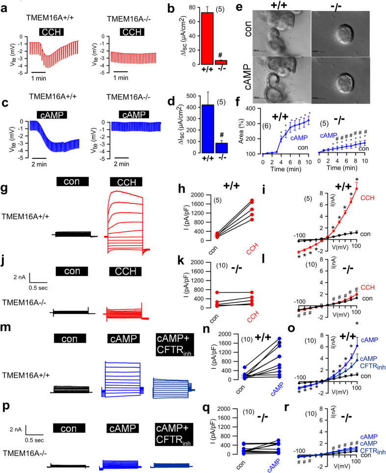
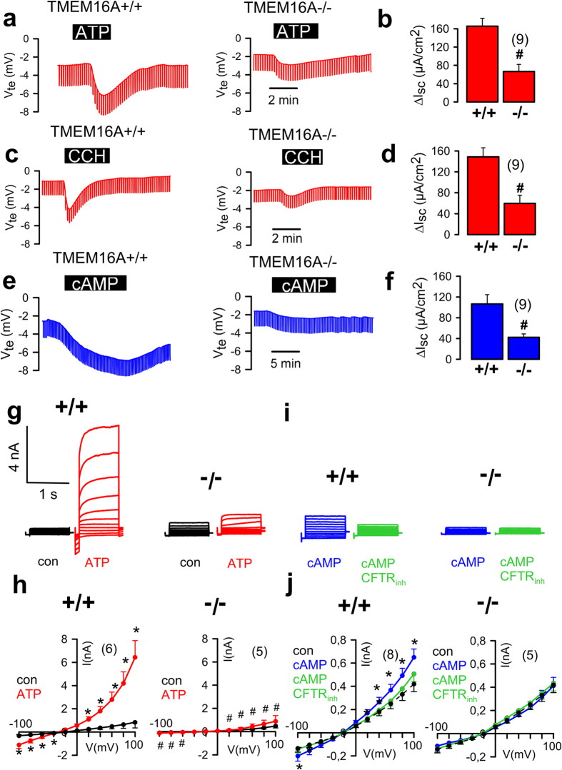
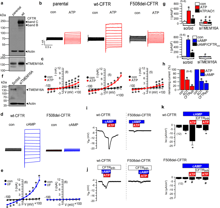
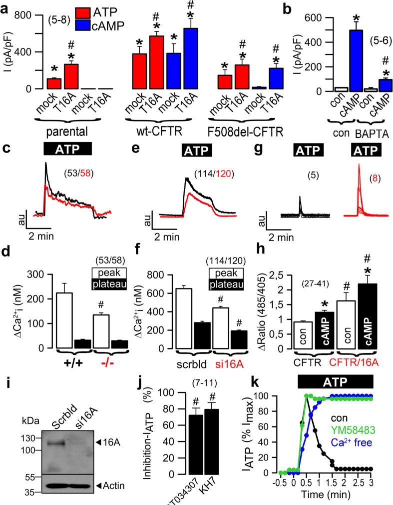
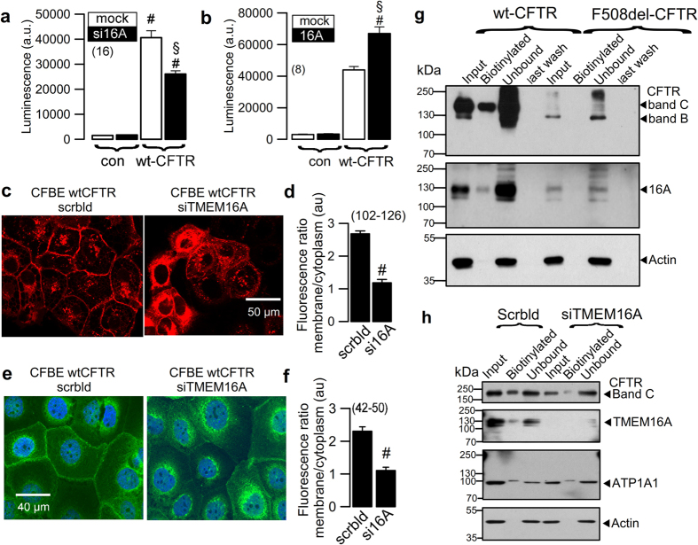
Cystic Fibrosis Transmembrane Conductance Regulator (CFTR) is the secretory chloride/bicarbonate channel in airways and intestine that is activated through ATP binding and phosphorylation by protein kinase A, but fails to operate in cystic fibrosis (CF). TMEM16A (also known as anoctamin 1, ANO1) is thought to function as the Ca2+ activated secretory chloride channel independent of CFTR. Here we report that tissue specific knockout of the TMEM16A gene in mouse intestine and airways not only eliminates Ca2+-activated Cl- currents, but unexpectedly also abrogates CFTR-mediated Cl- secretion and completely abolishes cAMP-activated whole cell currents. The data demonstrate fundamentally new roles of TMEM16A in differentiated epithelial cells: TMEM16A provides a mechanism for enhanced ER Ca2+ store release, possibly engaging Store Operated cAMP Signaling (SOcAMPS) and activating Ca2+ regulated adenylyl cyclases. TMEM16A is shown to be essential for proper activation and membrane expression of CFTR. This intimate regulatory relationship is the cause for the functional overlap of CFTR and Ca2+-dependent chloride transport.


Conflict of interest statement

The authors declare that they have no competing interests.

Figures

Figure 1
Figure 1
Intestinal epithelial knockout of TMEM16A eliminates CFTR currents. (a) Original recordings of the transepithelial voltage Vte and the effect of carbachol (CCH, 100 µM) in colonic epithelia from mice with intestinal epithelial knockout of TMEM16A (Vil1-Cre–TMEM16A flox/flox; TMEM16A−/−) and wild-type mice (TMEM16A+/+). (b) Summary of the calculated CCH-induced short circuit currents (ΔIsc). (c,d) Original recordings of Vte and summary of cAMP (IBMX 100 µM/forskolin 2 µM)-induced ΔIsc in +/+ and −/− colonic epithelia. (e,f) cAMP-induced secretion in intestinal organoids from +/+ and −/− intestines (upper panel) and summaries of luminal area increase (lower panel). (gi) Activation of whole cell currents by CCH and inhibition by CaCCinhAO1 (AO1; 10 µM) in isolated intestinal epithelial cells from +/+ mice. Original recordings (g), individual experiments (h) and current/voltage relationships (i). (jl) Corresponding experiments in intestinal epithelial cells from −/− mice. (mo) Activation of whole cell currents by cAMP and inhibition by CFTRinh172 (CFTRinh; 10 µM) in isolated intestinal epithelial cells from +/+ mice. (pr) Corresponding experiments in intestinal epithelial cells from −/− mice. Mean ± SEM; *Significant activation by cAMP or CCH (paired t-test). #Significant difference between −/− and +/+ (unpaired t-test). (number of mice or cells, respectively).
Figure 2
Figure 2
Respiratory epithelial knockout of TMEM16A eliminates CFTR currents. (a) Original recordings of the transepithelial voltage Vte and the effect of ATP (100 µM) in tracheas from FOXJ1-Cre–TMEM16A wt/wt (+/+) and FOXJ1-Cre–TMEM16A flox/flox (−/−) mice. (b) Summary of the calculated ATP-induced short circuit currents (ΔIsc). (c,d) Original recordings of Vte and summary of CCH-induced ΔIsc in +/+ and −/− tracheas. (e,f) Original recordings of Vte and summary of cAMP (IBMX 100 µM/forskolin 2 µM)-induced ΔIsc in +/+ and −/− tracheas. (g) Original recordings of whole cell currents activated by ATP in primary-cultured tracheal epithelial cells from TMEM16A+/+ and TMEM16A−/− mice. Experiments were performed in the presence of an inhibitor of Ca2+ activated K+ channels, TRAM-34 (100 nM). (h) Corresponding current/voltage relationships of whole cell currents activated by ATP in TMEM16A+/+ and TMEM16A−/− cells. (i) Original recordings of whole cell currents activated by cAMP in primary cultured respiratory epithelial cells from TMEM16A+/+ and TMEM16A−/− mice. (j) Corresponding current/voltage relationships of whole cell currents activated by cAMP in TMEM16A+/+ and TMEM16A−/− cells. Mean ± SEM; *Significant activation by cAMP or ATP (paired t-test). #Significant difference between −/− and +/+ (unpaired t-test). (number of mice or cells, respectively).
Figure 3
Figure 3
Cl currents by CFTR and TMEM16A in human airway epithelial cells cannot be strictly separated. (a) Western blot demonstrating wt-CFTR expression in CFBE/wt-CFTR cells, but not in CFBE parental cells. Both cells lines express similar levels of TMEM16A. (b) ATP (100 µM) -activated whole cell currents in CFBE parental cells and CFBE cells expressing wt-CFTR or F508del-CFTR. (c) Corresponding current/voltage relationships. (d) Whole cell currents activated by cAMP (100 µM IBMX/2 µM forskolin) in CFBE cells expressing wt-CFTR or F508del-CFTR. (e) Corresponding current/voltage relationships. (f) Western blot indicating suppression of TMEM16A in CFBE/wt-CFTR cells by siRNA (g) Summary of ATP (100 µM) -activated whole cell currents in CFBE/wt-CFTR cells treated with scrambled RNA or after siRNA-knockdown of TMEM16A (upper panel). Inhibition by CaCCinh-AO1 (10 µM). Summary of cAMP-activated whole cell currents in control (scrambled) and TMEM16A-knockdown cells, and effect of CFTRinh172 (10 µM). (h) Remaining currents after inhibition with CaCCinh_AO1 and CFTRinh172. Both blockers inhibit ATP- and cAMP-activated whole cell currents. (i) Transepithelial voltages recorded in polarized grown CFBE/wt-CFTR and CFBE/F508del-CFTR cells. Voltage deflections induced by cAMP or ATP. (j) Transepithelial voltages recorded in the presence of CFTRinh172 (10 µM). (k) Summaries of the corresponding calculated short circuit currents (Isc). Mean ± SEM; *Significant activation by ATP and cAMP, or currents inhibition by AO1 and CFTRinh, respectively (paired t-test). #Significant difference between scrambled and siTMEM16A or between wt-CFTR and F508del-CFTR, respectively (paired t-test). (number of cells).
Figure 4
Figure 4
TMEM16A provides Ca2+ for activation of CFTR. (a) Summary of whole cell currents activated by increase in intracellular Ca2+ (ATP; 100 µM) and cAMP (IBMX 100 µM/forskolin 2 µM) in parental cells, CFBE/wt-CFTR and CFBE/F508del-CFTR cells, with or without (mock) additional expression of exogenous TMEM16A. Additional TMEM16A augments ATP-induced currents in all cell lines, enhances cAMP-activated currents in CFBE/wt-CFTR cells, and induces cAMP-activated currents in CFBE/F508del-CFTR cells (b) cAMP-activated whole cell currents in CFBE/wt-CFTR cells were inhibited by the Ca2+ chelator BAPTA-AM. (c,d) Mean recordings of ATP-induced rise in intracellular Ca2+ (Fura2) in primary airway epithelial cells from TMEM16A+/+ (black) and TMEM16A−/− (red) mice (upper panel). Summary of peak and plateau Ca2+ increase (lower panel). (e,f) Mean recordings of ATP-induced rise in intracellular Ca2+ (Fura2) in CFBE/wt-CFTR cells (upper panel) and summary of peak and plateau Ca2+, which were reduced after siRNA-knockout (red) of TMEM16A (lower panel). (g,h) Recordings of ATP-induced Ca2+ peaks in HEK293 cells expressing GCAMP2-tagged CFTR. The ATP-induced Ca2+ peaks are larger in TMEM16A (red) coexpressing cells (upper panel). Summaries of Ca2+ peaks in the absence or presence of cAMP (lower panel). (i) Western blot indicating knockdown of TMEM16A expression by siRNA. (j) Inhibition (in %) of ATP-activated Cl currents by two different inhibitors of Ca2+-dependent adenylate cyclases, ST034307 (30 µM) and KH7 (10 µM). (k) Time courses for activation of whole cell currents by ATP (100 µM) under control conditions, in the presence of the ORAI-inhibitor YM58483, and in the absence of extracellular Ca2+. Mean ± SEM; *Significant activation by ATP or cAMP (paired t-test). #Significant difference when compared to mock, +/+, scrambled, absence of TMEM16A, or con, respectively (unpaired t-test). (number of cells or assays).
Figure 5
Figure 5
TMEM16A enhances membrane expression of CFTR. (a,b) Membrane expression of CFTR detected by chemiluminescence in CFBE/wt-CFTR cells expressing CFTR containing a FLAG tag in the first extracellular loop and a N-terminal cherry tag. Cells were exposed to a primary FLAG antibody (Sigma Taufkirchen, Germany, # F3165) and a secondary peroxidase-conjugated antibody. Luminescence was detected in CFBE/wt-CFTR cells but not in control parental cells (con). siRNA-knockdown of endogenous TMEM16A reduced chemiluminescence in CFBE/wt-CFTR cells (a), while additional expression of exogenous TMEM16A enhanced chemiluminescence (b). Very little background chemiluminescence was observed in the absence of CFTR (con). Mean ± SEM, (n) number of assays. #Significant difference when compared to mock (unpaired t-test). (c,d) Life imaging of cherry-CFTR in CFBE/wt-CFTR cells with and without siRNA-knockdown of TMEM16A, as detected by cherry fluorescence. Summary of fluorescence intensity ratios (plasma membrane/cytosolic fluorescence) indicating redistribution of the fluorescence towards cytoplasm by siRNA-TMEM16A. (e,f) Antibody-staining of CFTR in CFBE/wt-CFTR cells with and without siRNA-knockdown of TMEM16A. Summary of fluorescence intensity ratios (plasma membrane/cytosolic fluorescence) indicating redistribution of the fluorescence towards cytoplasm by siRNA-TMEM16A. (g) Membrane biotinylation of CFBE/wt-CFTR and CFBE/F508del-CFTR cells, and detection of membrane and cytosolic fractions of CFTR and TMEM16A using Western blot. (h) Effect of TMEM16A-knockdown on biotinylation (membrane expression) of CFTR and TMEM16A. Mean ± SEM; #Significant difference when compared to scrambled (paired t-test). (number of cells). Assays were performed in triplicates.
Figure 6
Figure 6
Molecular interaction of TMEM16A and CFTR. (a,b) Coimmunoprecipitation of CFTR and TMEM16A overexpressed in HEK293 cells. CFTR was pulled down by anti-CFTR antibody (Alomone labs, # ACL-006), while TMEM16A was pulled down by mouse monoclonal TMEM16A-antibody (Geneway GWB-MP178G). Both wt-CFTR and F508del-CFTR were found to interact with TMEM16A. Assays were performed in triplicates. (c,d) No coimmunoprecipitation was detected for CFTR and TMEM16F when experiments were performed under identical conditions using an TMEM16F antibody (Davids technology, Regensburg). White lines in the right blot indicate that lanes for unbound protein present on the same gel, have been relocated. Mean +/− SEM; #Indicates significant difference (p < 0.05; unpaired t-test). (number of experiments).

References

    1. Riordan JR. CFTR function and prospects for therapy. Annual review of biochemistry. 2008;77:701–726. doi: 10.1146/annurev.biochem.75.103004.142532. - DOI - PubMed
    1. Pedemonte N, Galietta LJ. Structure and Function of TMEM16 Proteins (Anoctamins) Physiol Rev. 2014;94:419–459. doi: 10.1152/physrev.00039.2011. - DOI - PubMed
    1. Kunzelmann K, Mall M. Electrolyte transport in the colon: Mechanisms and implications for disease. Physiological Reviews. 2002;82:245–289. doi: 10.1152/physrev.00026.2001. - DOI - PubMed
    1. Ousingsawat J, et al. Loss of TMEM16A causes a defect in epithelial Ca2+ dependent chloride transport. J Biol Chem. 2009;284:28698–28703. doi: 10.1074/jbc.M109.012120. - DOI - PMC - PubMed
    1. Huang F, et al. Calcium-activated chloride channel TMEM16A modulates mucin secretion and airway smooth muscle contraction. Proc. Natl. Acad. Sci USA. 2012;109:16354–16359. doi: 10.1073/pnas.1214596109. - DOI - PMC - PubMed

Publication types