. 2017 Sep 29;7(1):12409.
doi: 10.1038/s41598-017-12635-6.

Methylene blue inhibits NLRP3, NLRC4, AIM2, and non-canonical inflammasome activation

Affiliations

Methylene blue inhibits NLRP3, NLRC4, AIM2, and non-canonical inflammasome activation

Huijeong Ahn et al. Sci Rep. .

Abstract

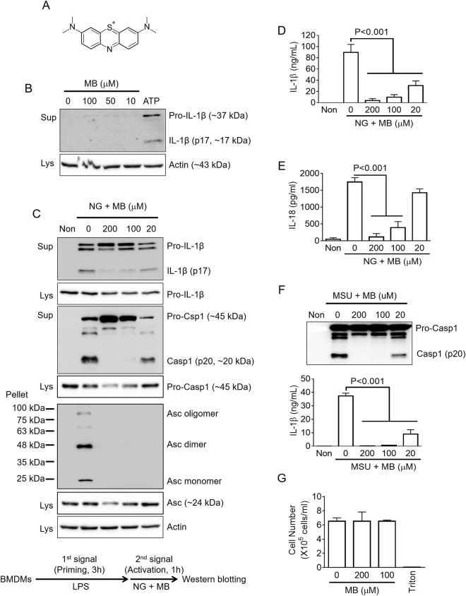
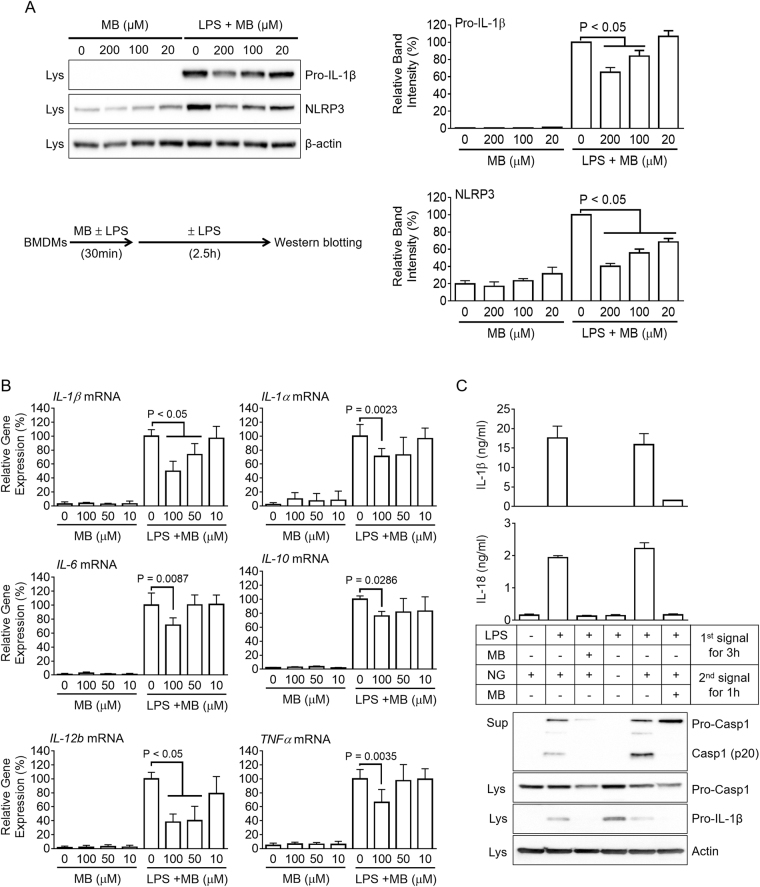
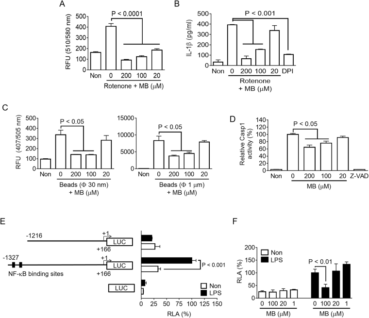
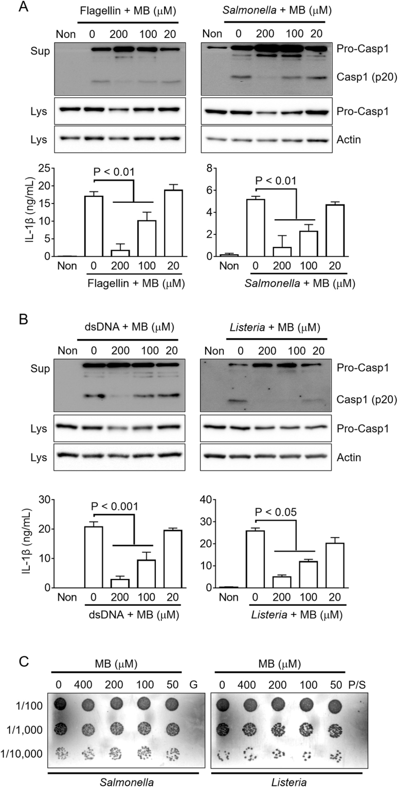
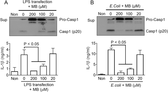
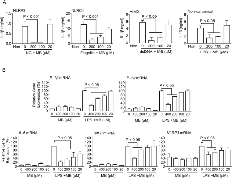
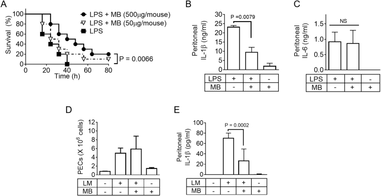
Methylene blue (MB), which has antioxidant, anti-inflammatory, neuroprotective, and mitochondria protective effects, has been widely used as a dye and medication. However, the effect of MB on inflammasome activation has not yet been studied. Inflammasomes are multi-protein complexes that induce maturation of interleukins (ILs)-1β and -18 as well as caspase-1-mediated cell death, known as pyroptosis. Dysregulation of inflammasomes causes several diseases such as type 2 diabetes, Alzheimer's disease, and gout. In this study, we assess the effect of MB on inflammasome activation in macrophages. As the result, MB attenuated activation of canonical inflammasomes such as NLRP3, NLRC4, and AIM2 as well as non-canonical inflammasome activation. In addition, MB inhibited upstream signals such as inflammasome assembly, phagocytosis, and gene expression of inflammasome components via inhibition of NF-κB signaling. Furthermore, MB reduced the activity of caspase-1. The anti-inflammasome properties of MB were further confirmed in mice models. Thus, we suggest that MB is a broad-spectrum anti-inflammasome candidate molecule.


Conflict of interest statement

The authors declare that they have no competing interests.

Figures

Figure 1
Figure 1
Effect of methylene blue on NLRP3 inflammasome activation. (A) Chemical structure of methylene blue (MB). (B) Lipopolysaccharide (LPS)-primed bone marrow-derived macrophages (LPS-primed BMDMs) were treated with the indicated concentration of MB or ATP (2 mM) as a positive control. Secretion of active form of IL-1β was analyzed by immunoblotting using cell culture supernatants (Sup) and cell lysates (Lys). (CE) LPS-primed BMDMs were treated with the indicated dosage of MB with/without nigericin (NG, 40 μM). (C) Secretion of IL-1β and caspase-1 (Casp1) and formation of Asc pyroptosome were analyzed by immunoblotting using Sup, Lys, and cross-linked pellets (Pellet) from whole cell lysates. The below schematic graph displays the chemical treatment process for inflammasome activation. IL-1β (D) and IL-18 (E) secretions were measured by ELISA. (F) LPS-primed BMDMs were treated with monosodium urate crystals (MSU, 800 μg/mL). Secretion of caspase-1 was analyzed by immunoblotting, and IL-1β secretion was measured by ELISA. (G) For cytotoxicity, BMDMs were treated the indicated dosages of MB, and cell number was measured by an automated cell counter. Triton x-100 (1%, Triton) treatment led to cell death. All immunoblot data shown are representative of at least three independent experiments. Bar graph presents the mean ± SD.
Figure 2
Figure 2
Effect of MB on the priming step of inflammasome activation and expression of other cytokine genes. (A and B) BMDMs were treated with the indicated concentration of MB with/without LPS (10 ng/mL) as indicated in the schematic graph. (A) Pro-IL-1β and NLRP3 expression levels were analyzed by immunoblotting and further presented with band density. (B) Expression levels of mouse IL-1β, IL-1α, IL-6, IL-10, IL-12b, and TNFα mRNAs were quantitated by real-time PCR. C, BMDMs were treated with MB and/or LPS as the 1st signal, after which cells were replaced by media containing nigericin (NG, 2nd signal) with/without MB as the 2nd signal. IL-1β and IL-18 secretion levels were measured by ELISA, and Casp1 secretion and pro-IL1β expression were analyzed by immunoblotting. All immunoblot data shown are representative of at least three independent experiments. Bar graph presents the mean ± SD.
Figure 3
Figure 3
Effect of MB on mitochondrial ROS production, phagocytosis, caspase-1 activity, and NLRP3 promoter activity. (A) BMDMs were treated with rotenone (160 μM) and the indicated dosages of MB for 6 h, after which mitochondrial ROS generation was analyzed. (B) LPS-primed BMDMs were treated with rotenone with/without MB, and secretion of IL-1β was measured by ELISA. (C) BMDMs were incubated with two different diameters (30 nm or 1 μm) of fluorescent beads for 6 h, followed by measurement of intracellular beads based on florescence intensity. (D) Recombinant human caspase-1 (rhCasp1) was incubated with its substrate (YVAD-pNA) in the presence of MB as indicated. (E) Luciferase-expressing plasmids (pGL3) controlled by mouse NLRP3 promoters (−1,327 nt to + 166 nt or −1,216 nt to + 166 nt) were transfected into RAW 264.7 cells, which were treated with/without LPS. Left schematic figures indicate the mouse NLRP3 promoter regions (filled boxes, NF-κB binding sites; LUC, luciferase gene). (F) RAW 264.7 cells were transfected with pGL3 (−1,327 nt to + 166 nt) and treated with MB and/or LPS. Relative luciferase activity (RLA) was analyzed. Bar graph presents the mean ± SD.
Figure 4
Figure 4
Effect of MB on NLRC4 or AIM2 inflammasome activation. (A) For NLRC3 inflammasome activation, LPS-primed BMDMs were treated with the indicated dosage of MB in the presence of flagellin (0.5 μg/mL) or Salmonella typhimurium (MOI 3.5) for 1 h. (B) For AIM2 inflammasome activation, macrophages were primed with LPS and treated with MB and dsDNA (1 μg/mL) for 1 h or Listeria monocytogenes (MOI 35) for 3 h. Secretions of caspase-1 (Casp1) and IL-1β were measured by immunoblotting and ELISA. (C) For bactericidal properties of MB, diluted Salmonella or Listeria were cultured in LB or BHI plates with the indicated concentration of MB or antibiotics. G gentamycin (50 μg/mL); P/S, penicillin (100 I.U.), and streptomycin (100 μg/mL). All immunoblot and bacterial growing data shown are representative of at least three independent experiments. Bar graph presents the mean ± SD.
Figure 5
Figure 5
Effect of MB on non-canonical inflammasome activation. (A) LPS-primed BMDMs were transfected with LPS for non-canonical inflammasome activation in the presence of MB as indicated. (B) LPS-primed BMDMs were infected with E.coli (MOI = 10) in the presence of MB as indicated. Cspase-1 (Casp1) and IL-1β secretions were analyzed by immunoblotting and ELISA. All immunoblot data shown are representative of at least three independent experiments. Bar graph presents the mean ± SD.
Figure 6
Figure 6
Effect of MB on inflammasome activation in a human monocyte-like cell line, THP-1. (A) PMA-treated THP-1 cells were primed with LPS and then subjected to NG treatment and transfection of flagellin, dsDNA, and LPS in order to activate NLRP3, NLRC4, AIM2, and non-canonical inflammasomes. Secretion of IL-1β was analyzed by ELISA. (B) Expression levels of human IL-1β, IL-1α, IL-6, TNFα, and NLRP3 mRNAs were quantitated by real-time PCR. Bar graph presents the mean ± SD.
Figure 7
Figure 7
Effect of MB on inflammasome model animals and non-canonical inflammasome. (A) Mice (n = 10 per group) were intraperitoneally (ip) injected with LPS (25 mg/Kg) and/or MB as indicated. Survival rates were observed at the indicated times. (B and C), Mice (n = 5 per group) were ip-injected with LPS (4 mg/kg) and/or MB (500 μg/mouse). IL-1β (B) and IL-6 (C) secretion levels in peritoneal lavage fluids were measured. (D and E), Mice (n = 9 per group) were ip injected with Listeria monocytogenes (1,000 cfu in 200 μL of saline) and/or MB (500 μg/mouse). Number of peritoneal exudate cells (PECs, B) was calculated, and IL-1β (C) secretion levels of peritoneal lavage fluids were measured. Bar graph presents the mean ± SD.

Similar articles

Cited by

References

    1. Schirmer RH, Adler H, Pickhardt M, Mandelkow E. Lest we forget you–methylene blue. Neurobiology of aging. 2011;32:2325 e2327–2316. doi: 10.1016/j.neurobiolaging.2010.12.012. - DOI - PubMed
    1. Oz M, Lorke DE, Petroianu GA. Methylene blue and Alzheimer’s disease. Biochemical pharmacology. 2009;78:927–932. doi: 10.1016/j.bcp.2009.04.034. - DOI - PubMed
    1. Fenn AM, et al. Methylene blue attenuates traumatic brain injury-associated neuroinflammation and acute depressive-like behavior in mice. J Neurotrauma. 2015;32:127–138. doi: 10.1089/neu.2014.3514. - DOI - PMC - PubMed
    1. Chen GY, Nunez G. Sterile inflammation: sensing and reacting to damage. Nat Rev Immunol. 2010;10:826–837. doi: 10.1038/nri2873. - DOI - PMC - PubMed
    1. Guo H, Callaway JB, Ting JP. Inflammasomes: mechanism of action, role in disease, and therapeutics. Nat Med. 2015;21:677–687. doi: 10.1038/nm.3893. - DOI - PMC - PubMed

Publication types

MeSH terms