. 2017 Sep 29;7(1):12423.
doi: 10.1038/s41598-017-12403-6.

Radiation alters the cargo of exosomes released from squamous head and neck cancer cells to promote migration of recipient cells

Affiliations

Radiation alters the cargo of exosomes released from squamous head and neck cancer cells to promote migration of recipient cells

Lisa Mutschelknaus et al. Sci Rep. .

Abstract

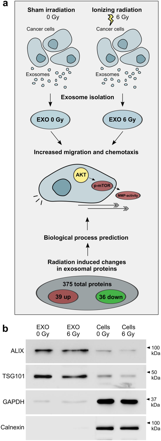
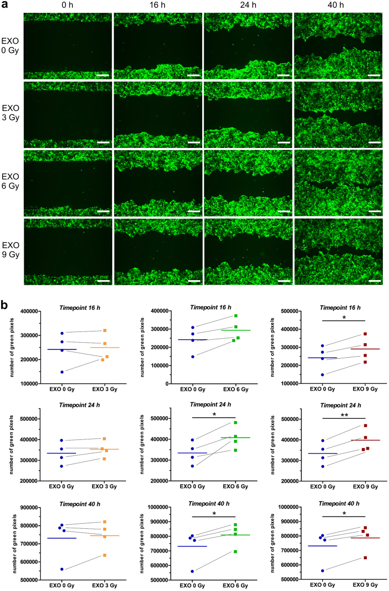
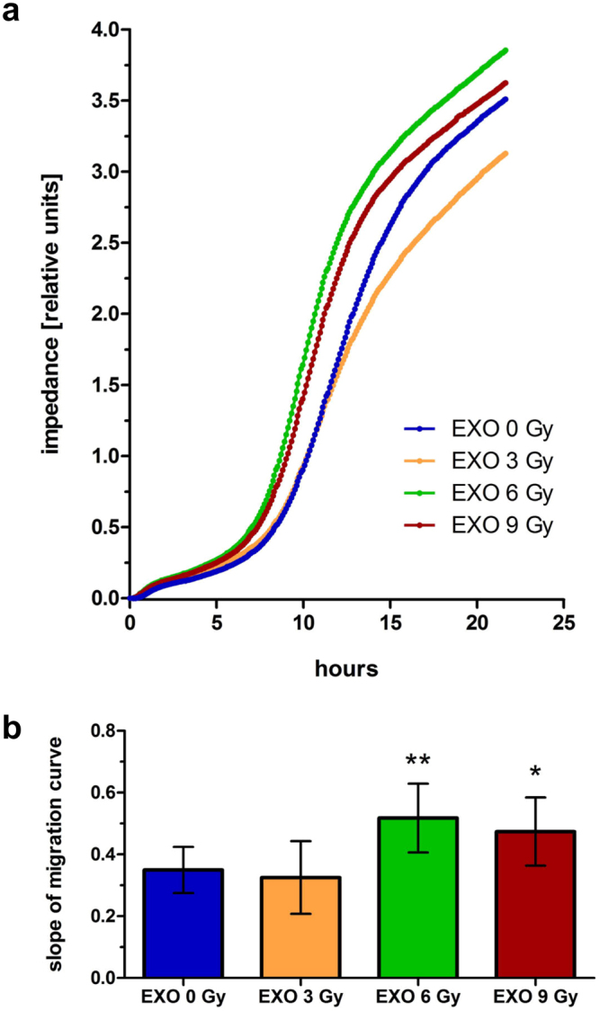
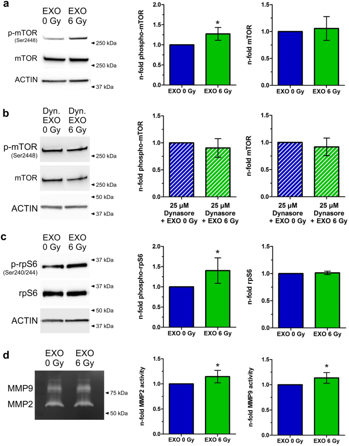
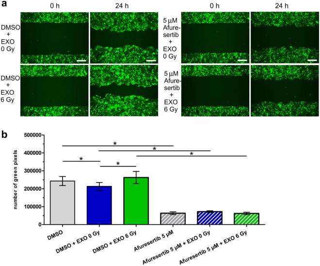
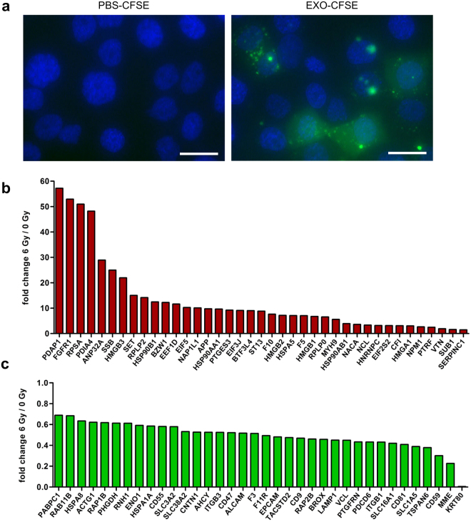
Radiation is a highly efficient therapy in squamous head and neck carcinoma (HNSCC) treatment. However, local recurrence and metastasis are common complications. Recent evidence shows that cancer-cell-derived exosomes modify tumour cell movement and metastasis. In this study, we link radiation-induced changes of exosomes to their ability to promote migration of recipient HNSCC cells. We demonstrate that exosomes isolated from irradiated donor cells boost the motility of the HNSCC cells BHY and FaDu. Molecular data identified enhanced AKT-signalling, manifested through increased phospho-mTOR, phospho-rpS6 and MMP2/9 protease activity, as underlying mechanism. AKT-inhibition blocked the pro-migratory action, suggesting AKT-signalling as key player in exosome-mediated migration. Proteomic analysis of exosomes isolated from irradiated and non-irradiated BHY donor cells identified 39 up- and 36 downregulated proteins. In line with the observed pro-migratory effect of exosomes isolated from irradiated cells protein function analysis assigned the deregulated exosomal proteins to cell motility and AKT-signalling. Together, our findings demonstrate that exosomes derived from irradiated HNSCC cells confer a migratory phenotype to recipient cancer cells. This is possibly due to radiation-regulated exosomal proteins that increase AKT-signalling. We conclude that exosomes may act as driver of HNSCC progression during radiotherapy and are therefore attractive targets to improve radiation therapy strategies.


Conflict of interest statement

The authors declare that they have no competing interests.

Figures

Figure 1
Figure 1
Functional and molecular comparison of exosomes released from 6 Gy irradiated and non-irradiated head and neck cancer cells. Exosomes isolated from irradiated BHY cells induce migration and chemotaxis by activating AKT-signalling and extracellular MMPs. In the same line radiation-induced changes of exosomal proteins predict effects on migration, chemotaxis and AKT-signalling. (b) Representative, cropped western blot of exosome markers ALIX and TSG101 as well as cytosolic markers GAPDH and Calnexin for BHY exosomes and cells isolated 24 hours after 0 and 6 Gy irradiation.
Figure 2
Figure 2
Exosomes from irradiated BHY cells enhance the migratory phenotype. (a) Exemplary wound healing of BHY-GFP cells after 16, 24 and 40 hours (scale bar: 500 µm). Cells were either preincubated with exosomes from non-irradiated (EXO 0 Gy), 3 Gy (EXO 3 Gy), 6 Gy (EXO 6 Gy) or 9 Gy (EXO 9 Gy) irradiated BHY cells. (b) Quantification of the wound healing capacity with the Image Colour Analyser after 16, 24 and 40 hours [n = 4; two-sided, paired t-test; p-value < 0.05].
Figure 3
Figure 3
Exosomes from irradiated BHY cells enhance the chemotaxis-induced motility. The xCELLigence system was used to analyse the chemotactic movement of cells after a 24 hours pretreatment with exosomes from non-irradiated (EXO 0 Gy), 3 Gy (EXO 3 Gy), 6 Gy (EXO 6 Gy) or 9 Gy (EXO 9 Gy) irradiated BHY cells. (a) Mean impedance as measure of transfilter migration of cells is plotted over time. (b) Slope of the migration curves [n = 3; ± SD; two-sided, unpaired t-test; *p-value < 0.05; **p-value < 0.01].
Figure 4
Figure 4
Exosomes from irradiated cells activate the AKT-pathway. (a) Western blot of phospho-mTOR (Ser2448) and mTOR of cells which were incubated for 24 hours with exosomes isolated either from irradiated cells (EXO 6 Gy) or from non-irradiated cells (EXO 0 Gy). Normalization was performed to ACTIN and to cells treated with exosomes from non-irradiated cells (EXO 0 Gy). Cropped blots are displayed [n = 4; ± SD; two-sided, one-sample t-test; p-value < 0.05]. (b) Western blot of phospho-mTOR (Ser2448) and mTOR of cells which were pretreated for 1 hour with 25 µM Dynasore and incubated for 24 hours with exosomes isolated either from irradiated cells (EXO 6 Gy) or from non-irradiated cells (EXO 0 Gy). Normalization was performed to ACTIN and to cells treated with exosomes from non-irradiated cells (EXO 0 Gy). Cropped blots are displayed [n = 3; ±SD; two-sided, one-sample t-test]. (c) Western blot of phospho-S6 Ribosomal Protein (Ser240/244) and S6 Ribosomal Protein of cells which were incubated for 24 hours with exosomes isolated either from irradiated cells (EXO 6 Gy) or from non-irradiated cells (EXO 0 Gy). Normalization was performed to ACTIN and to cells treated with exosomes from non-irradiated cells (EXO 0 Gy). Cropped blots are displayed [n = 7; ±SD; two-sided, one-sample t-test; p-value < 0.05]. (d) MMP2 and MMP9 activity in the supernatants 24 hours after transfer of exosomes isolated from irradiated (EXO 6 Gy) and from non-irradiated cells (EXO 0 Gy) on BHY cells. Normalization was performed to cells treated with EXO 0 Gy. Cropped gels are displayed [n = 6; ±SD; two-sided, one-sample t-test; p-value < 0.05].
Figure 5
Figure 5
Exosomes from irradiated cells activate the AKT-pathway to induce migration. (a) Exemplary wound healing of BHY-GFP cells after treatment with 5 µM of the AKT-inhibitor Afuresertib or DMSO, in combination with exosomes isolated from irradiated (EXO 6 Gy) and from non-irradiated cells (EXO 0 Gy). The pictures were taken 24 hours after migration start (scale bar: 500 µm). (b) Quantification of wound healing capacity with the Image Colour Analyser 24 hours [n = 3; ±SD; two-sided, paired t-test; p-value < 0.05].
Figure 6
Figure 6
Exosomes from head and neck cancer cells transfer proteins to recipient cells and have a modified protein composition after ionizing radiation. (a) Exosomal proteins (EXO-CFSE) of BHY cells and PBS (PBS-CFSE) as negative control were stained with CFSE and subsequently transferred onto recipient BHY cells. The protein uptake was monitored after 24 hours of exposure (scale bar: 25 µm). Protein analysis of exosomes isolated 24 hours after 6 Gy irradiation of the head and neck cancer cell line BHY revealed (b) 39 upregulated and (c) 36 downregulated proteins [n = 3; FDR-adjusted p-value (q-value), ≥2 unique peptides, fold-change between ≤0.7 and ≥1.3; q-value < 0.05.

References

    1. Neville BW, Day TA. Oral cancer and precancerous lesions. CA Cancer J Clin. 2002;52:195–215. doi: 10.3322/canjclin.52.4.195. - DOI - PubMed
    1. Vilalta M, Rafat M, Graves EE. Effects of radiation on metastasis and tumor cell migration. Cellular and molecular life sciences: CMLS. 2016;73:2999–3007. doi: 10.1007/s00018-016-2210-5. - DOI - PMC - PubMed
    1. Moncharmont C, et al. Radiation-enhanced cell migration/invasion process: a review. Crit Rev Oncol Hematol. 2014;92:133–142. doi: 10.1016/j.critrevonc.2014.05.006. - DOI - PubMed
    1. Edalat L, et al. BK K+ channel blockade inhibits radiation-induced migration/brain infiltration of glioblastoma cells. Oncotarget. 2016;7:14259–14278. doi: 10.18632/oncotarget.7423. - DOI - PMC - PubMed
    1. Strong MS, et al. A randomized trial of preoperative radiotherapy in cancer of the oropharynx and hypopharynx. Am J Surg. 1978;136:494–500. doi: 10.1016/0002-9610(78)90268-4. - DOI - PubMed

MeSH terms