

Squamous cell carcinoma of the lung showing a ground glass nodule on high-resolution computed tomography associated with pneumoconiosis: a case report
- PMID: 28963659
- PMCID: PMC5622012
- DOI: 10.1186/s40792-017-0384-1
Squamous cell carcinoma of the lung showing a ground glass nodule on high-resolution computed tomography associated with pneumoconiosis: a case report
Abstract
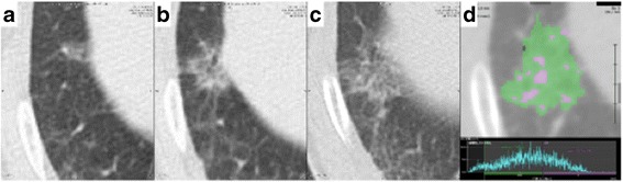
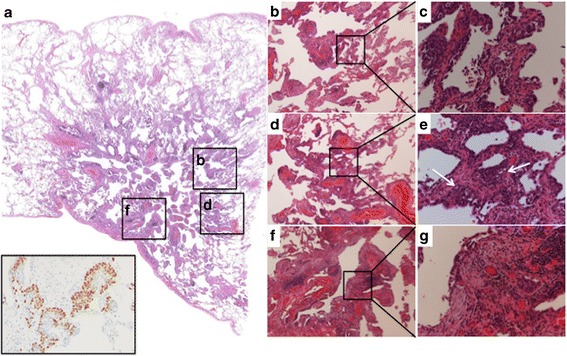
Background: Adenocarcinoma with lepidic growth pattern presents as a ground glass nodule (GGN) on high resolution computed tomography (CT), whereas peripheral pulmonary squamous cell carcinoma (SCC) usually presents as a solid nodule. We herein report a rare case of pulmonary SCC extending along the alveolar lumen representing as a GGN on a CT scan in a patient with pneumoconiosis.
Case presentation: A 77-year-old man with pneumoconiosis was found to have a gradually enlarging GGN in the right lower lobe of the lung on CT. An adenocarcinoma of the lung was suspected. The GGN was successfully resected by thoracoscopic segmentectomy. Pathological examination of the resected specimen was pathologically diagnosed as a stage IA SCC extending along the alveolar lumen. The patient had no evidence of recurrence 19 months after surgery.
Conclusions: SCC should be included in the differential diagnosis of peripherally located GGNs, especially in patients at high risk of SCC of the lung such as those with pneumoconiosis.
Keywords: Ground glass nodule; Lepidic growth; Lung cancer; Pneumoconiosis; Squamous cell carcinoma.
Conflict of interest statement
Consent for publication
Written informed consent was obtained from the patient for the publication of this case report and the accompanying images.
Competing interests
The authors declare that they have no competing interests.
Publisher’s Note
Springer Nature remains neutral with regard to jurisdictional claims in published maps and institutional affiliations.
Figures
References
-
- Atsumi J, Shimizu K, Kakegawa S, et al. A case of squamous cell carcinoma spreading along the alveolar walls with a ground glass opacity on high resolution computed tomography (HRCT) Haigan. 2010;50(4):379–380. doi: 10.2482/haigan.50.379. - DOI
-
- Kimura T, Asano F, Wakahara K, et al. Early image of squamous cell carcinoma in the peripheral lung field on high-resolution CT scan. Nihon Kokyuki Gakkai Zasshi. 2002;40(11):884–888. - PubMed
LinkOut - more resources
Full Text Sources
Other Literature Sources
Research Materials
