Prion-like transmission of α-synuclein pathology in the context of an NFL null background
- PMID: 28964772
- PMCID: PMC5813496
- DOI: 10.1016/j.neulet.2017.09.054
Prion-like transmission of α-synuclein pathology in the context of an NFL null background
Abstract
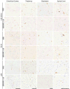
Neurofilaments are a major component of the axonal cytoskeleton in neurons and have been implicated in a number of neurodegenerative diseases due to their presence within characteristic pathological inclusions. Their contributions to these diseases are not yet fully understood, but previous studies investigated the effects of ablating the obligate subunit of neurofilaments, low molecular mass neurofilament subunit (NFL), on disease phenotypes in transgenic mouse models of Alzheimer's disease and tauopathy. Here, we tested the effects of ablating NFL in α-synuclein M83 transgenic mice expressing the human pathogenic A53T mutation, by breeding them onto an NFL null background. The induction and spread of α-synuclein inclusion pathology was triggered by the injection of preformed α-synuclein fibrils into the gastrocnemius muscle or hippocampus in M83 versus M83/NFL null mice. We observed no difference in the post-injection time to motor-impairment and paralysis endpoint or amount and distribution of α-synuclein inclusion pathology in the muscle injected M83 and M83/NFL null mice. Hippocampal injected M83/NFL null mice displayed subtle region-specific differences in the amount of α-synuclein inclusions however, pathology was observed in the same regions as the M83 mice. Overall, we observed only minor differences in the induction and transmission of α-synuclein pathology in these induced models of synucleinopathy in the presence or absence of NFL. This suggests that NFL and neurofilaments do not play a major role in influencing the induction and transmission of α-synuclein aggregation.
Keywords: NFL gene (NEFL) knockout; Neurofilaments; Parkinson’s disease; Synucleinopathy; Transgenic mice; α-Synuclein.
Copyright © 2017 Elsevier B.V. All rights reserved.
Conflict of interest statement
Conflicts of interest: none.
Figures


Similar articles
-
Comparative analyses of the in vivo induction and transmission of α-synuclein pathology in transgenic mice by MSA brain lysate and recombinant α-synuclein fibrils.Acta Neuropathol Commun. 2019 May 20;7(1):80. doi: 10.1186/s40478-019-0733-3. Acta Neuropathol Commun. 2019. PMID: 31109378 Free PMC article.
-
Robust Central Nervous System Pathology in Transgenic Mice following Peripheral Injection of α-Synuclein Fibrils.J Virol. 2017 Jan 3;91(2):e02095-16. doi: 10.1128/JVI.02095-16. Print 2017 Jan 15. J Virol. 2017. PMID: 27852849 Free PMC article.
-
Comparison of the in vivo induction and transmission of α-synuclein pathology by mutant α-synuclein fibril seeds in transgenic mice.Hum Mol Genet. 2017 Dec 15;26(24):4906-4915. doi: 10.1093/hmg/ddx371. Hum Mol Genet. 2017. PMID: 29036344 Free PMC article.
-
[Prion-like Propagation of Pathological α-Synuclein in Vivo].Yakugaku Zasshi. 2019;139(7):1007-1013. doi: 10.1248/yakushi.18-00165-4. Yakugaku Zasshi. 2019. PMID: 31257247 Review. Japanese.
-
The concept of alpha-synuclein as a prion-like protein: ten years after.Cell Tissue Res. 2018 Jul;373(1):161-173. doi: 10.1007/s00441-018-2814-1. Epub 2018 Feb 26. Cell Tissue Res. 2018. PMID: 29480459 Free PMC article. Review.
Cited by
-
Differential gene expression and immune profiling in Parkinson's disease: unveiling potential candidate biomarkers.BMC Neurol. 2025 Aug 27;25(1):354. doi: 10.1186/s12883-025-04388-x. BMC Neurol. 2025. PMID: 40859216 Free PMC article.
-
Comparative analyses of the in vivo induction and transmission of α-synuclein pathology in transgenic mice by MSA brain lysate and recombinant α-synuclein fibrils.Acta Neuropathol Commun. 2019 May 20;7(1):80. doi: 10.1186/s40478-019-0733-3. Acta Neuropathol Commun. 2019. PMID: 31109378 Free PMC article.
References
-
- Julien JP, Mushynski WE. Neurofilaments in health and disease. Prog Nucleic Acid Res Mol Biol. 1998;61:1–23. - PubMed
MeSH terms
Substances
Grants and funding
LinkOut - more resources
Full Text Sources
Other Literature Sources
Molecular Biology Databases

