. 2017 Dec 28:411:35-43.
doi: 10.1016/j.canlet.2017.09.032. Epub 2017 Sep 28.

Synergistic efficacy of irinotecan and sunitinib combination in preclinical models of anaplastic thyroid cancer

Affiliations

Synergistic efficacy of irinotecan and sunitinib combination in preclinical models of anaplastic thyroid cancer

Teresa Di Desidero et al. Cancer Lett. .

Abstract

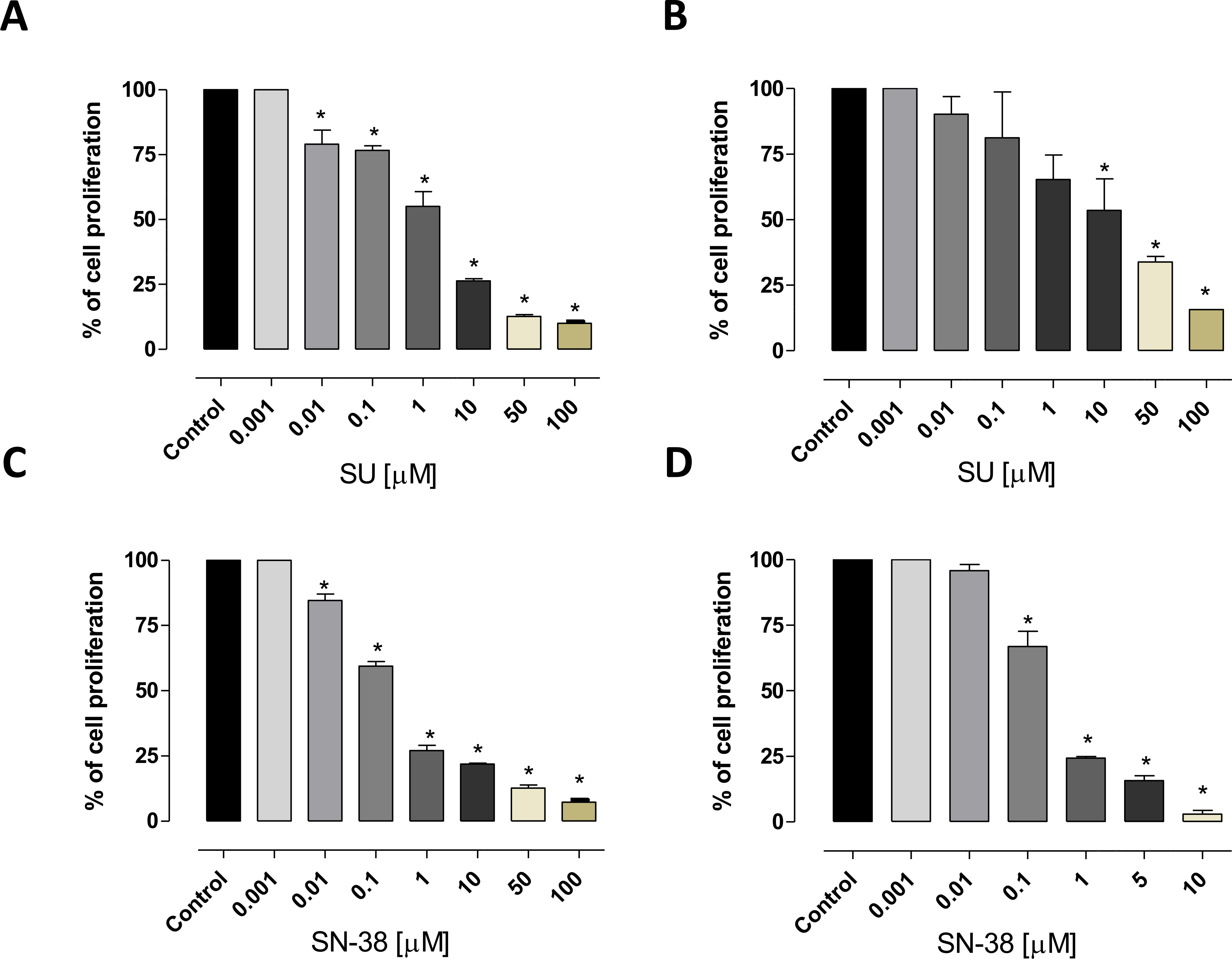
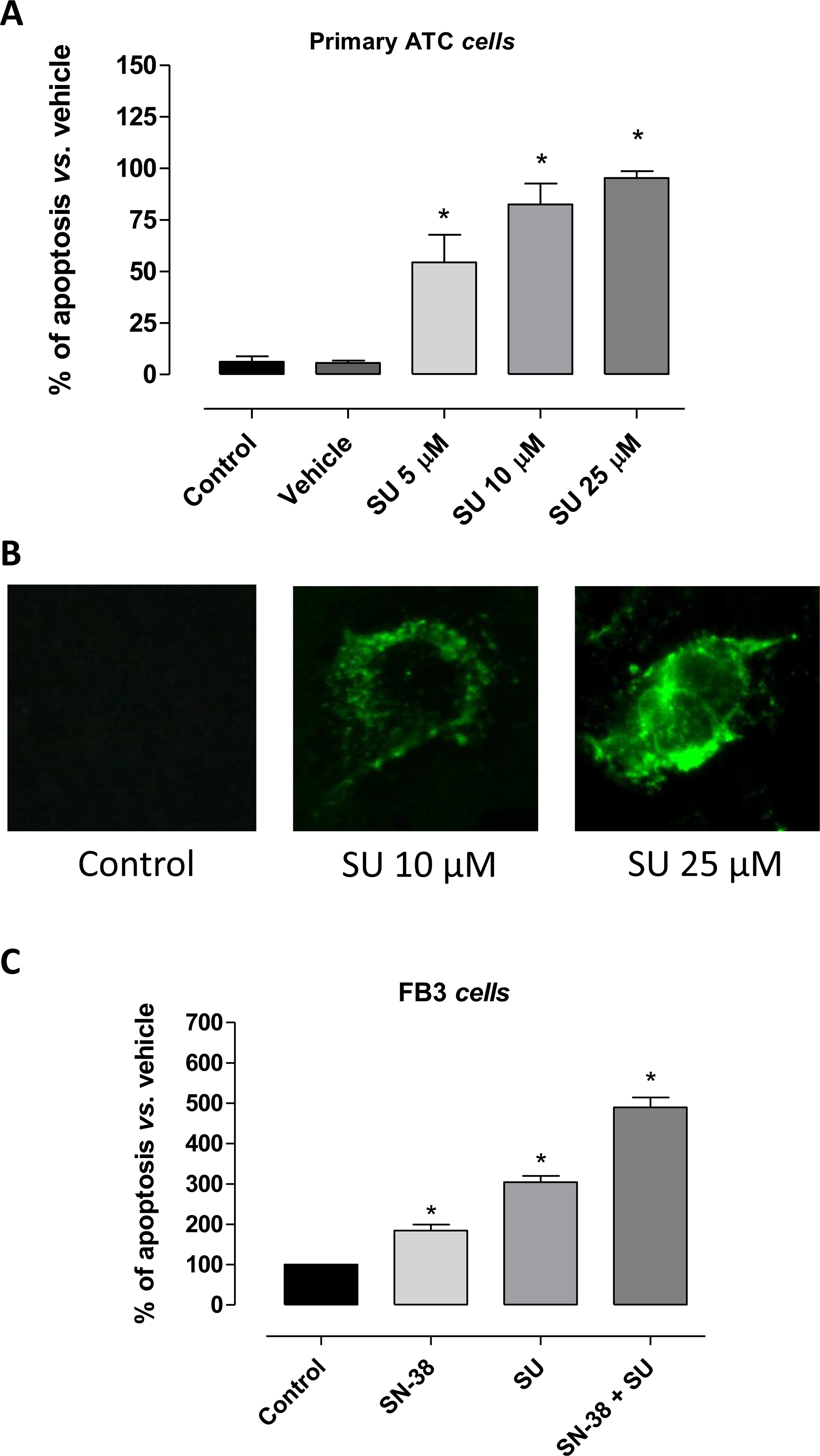
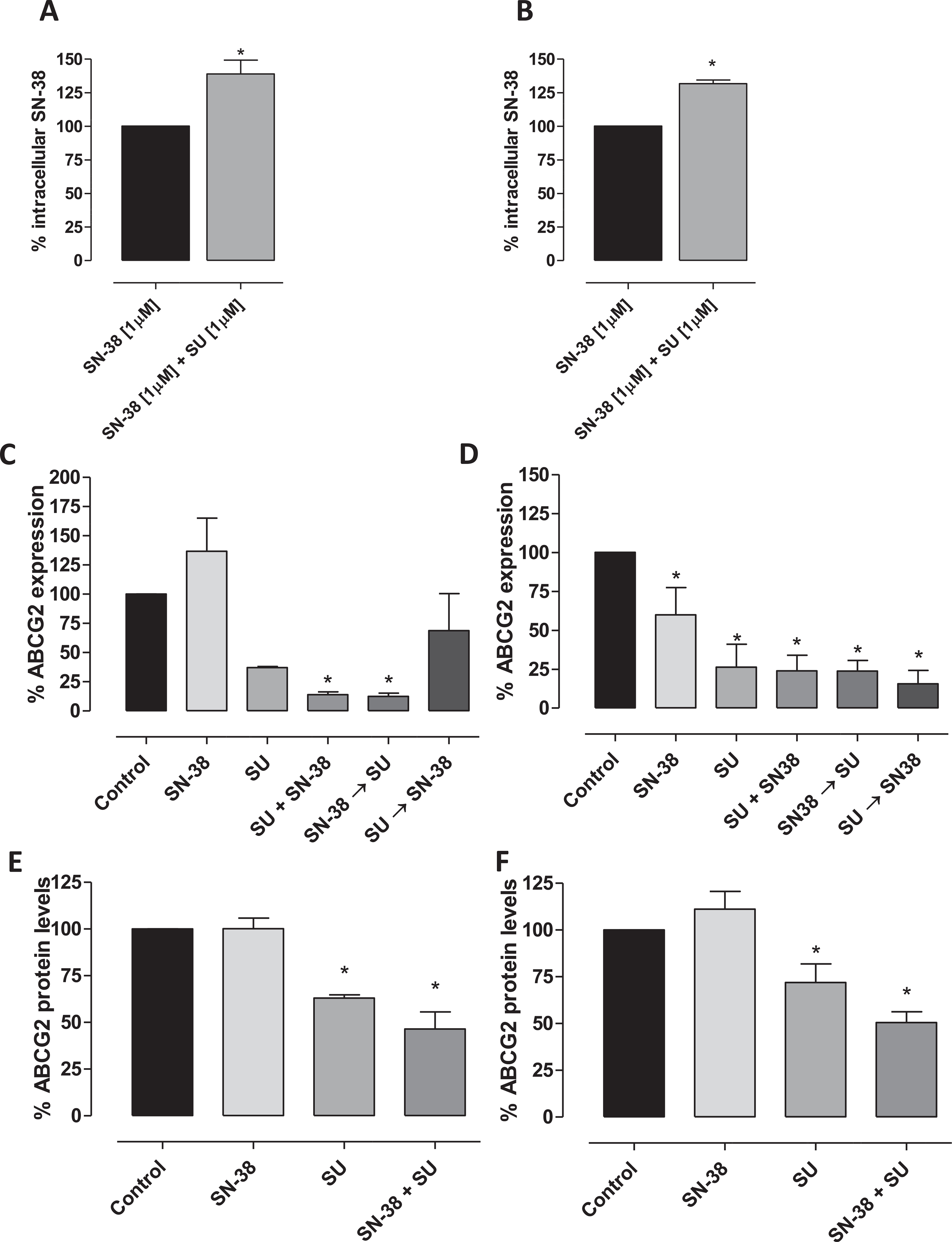
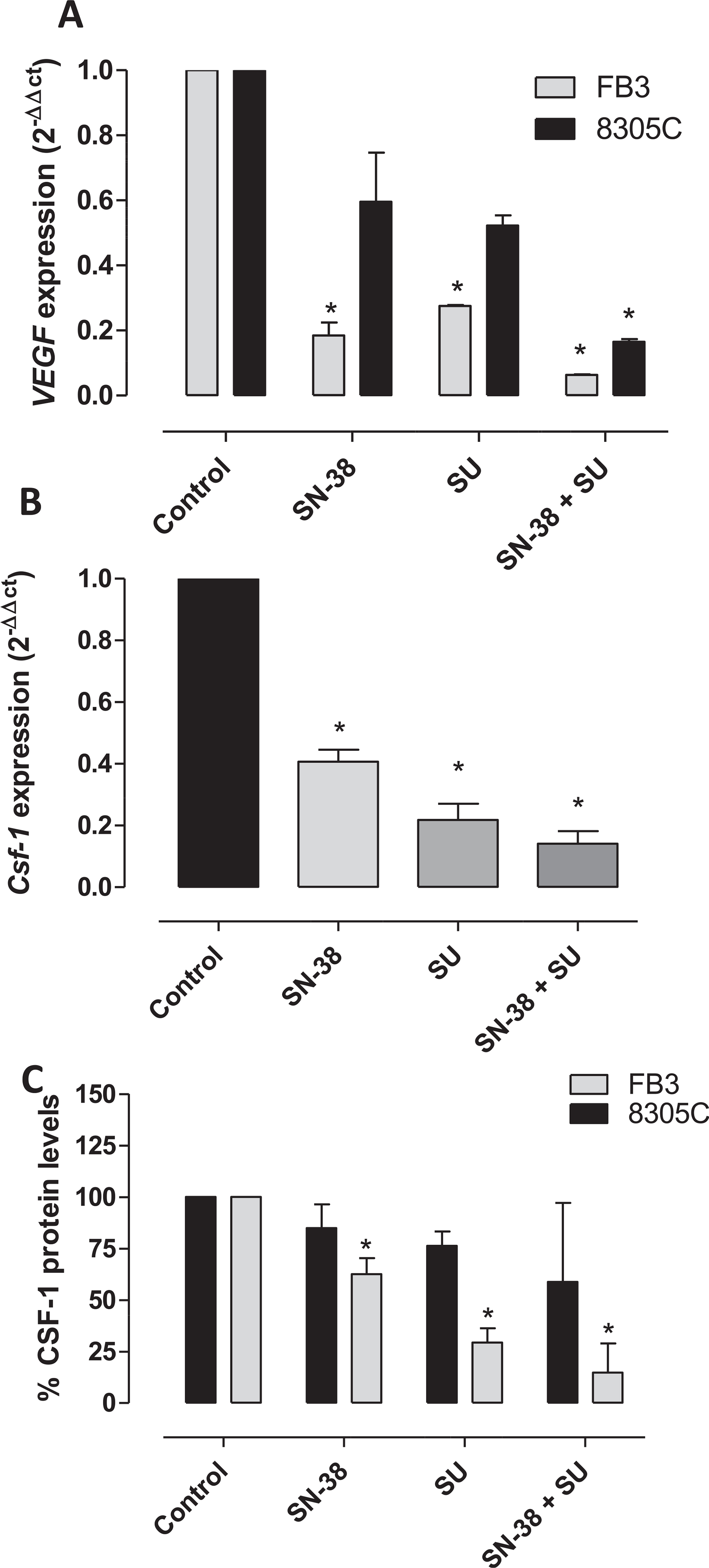
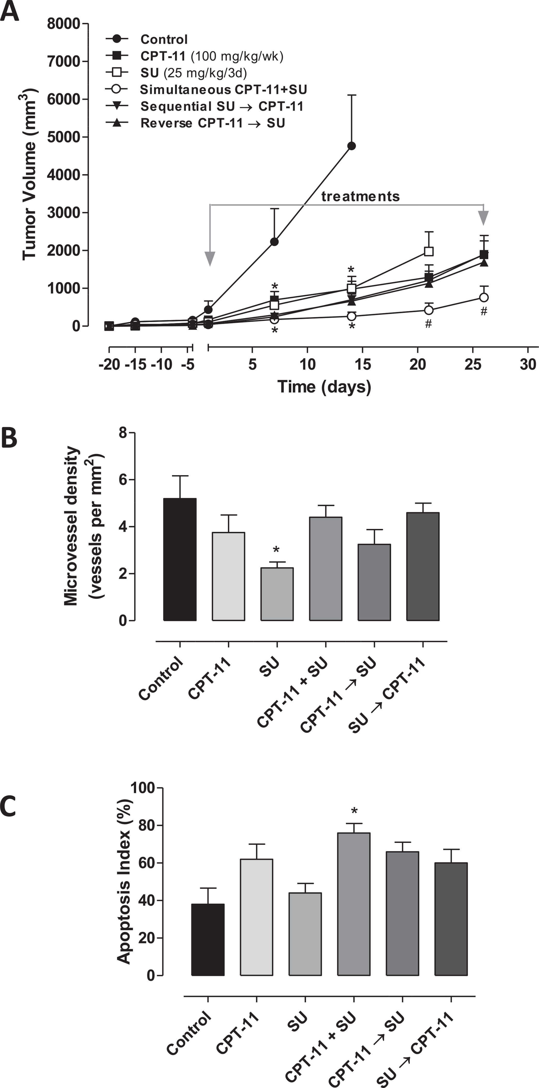
The identification of new therapeutic strategies is urgently needed for the management of patients affected by anaplastic thyroid cancer (ATC) due to their short survival and poor prognosis. Aim of the study was to determine the activity of the combination irinotecan/sunitinib on ATC cell growth in vitro and the antitumor effects in vivo. Proliferation assays were performed for 72 h on ATC cell lines exposed to the combination of SN-38, the active metabolite of irinotecan, and sunitinib. The simultaneous combination of sunitinib and SN-38, quantified by the combination index, determined a high synergism on ATC cells, increasing the intracellular concentrations of SN-38. Moreover, the synergistic combination greatly decreases the gene expression and the protein levels of vascular endothelial growth factor, colony stimulating factor 1 and ATP-binding cassette transporter G2 in ATC cells. A significant in vivo antitumor effect was observed in ATC xenografts with the simultaneous combination of irinotecan and sunitinib if compared to monotherapy. The simultaneous combination of irinotecan and sunitinib, in vitro and in vivo demonstrated a significant, synergistic ATC antitumor activity, suggesting a possible and rapid translation of this schedule into the clinics.

Keywords: Anaplastic thyroid cancer; Irinotecan; Sunitinib; Synergism; Tumor xenografts.


Conflict of interest statement

Conflict of interest

None.

Figures

Fig. 1.
Fig. 1.
Antiproliferative effects of sunitinib (SU) and SN-38 in vitro on 8305C (A and C, respectively), and FB3 (B and D, respectively) cell lines. The antiproliferative effects of the drugs were studied after 72 h of exposure. The data are presented as percentage of vehicle-treated cells. The concentrations of drug that reduced cell proliferation by 50% (IC50) vs controls were calculated by a nonlinear regression fit of the mean values of the data obtained in triplicate experiments (i.e. at least 9 wells for each concentration). Columns and bars, mean values ± S.E., respectively. *, P < 0.001 vs. control.
Fig. 2.
Fig. 2.
A) Apoptosis in primary ATC cells treated with sunitinib (SU) for 24 h (mean ± SD n = 5). Data were analyzed by one-way ANOVA with Newman–Keuls multiple comparisons test and with a test for linear trend (*P < 0.001 vs. control). The percentage of apoptotic cells after the treatment with vehicle (DMSO) was not significantly different from the one of control (not treated cells). B) Representative images of immunofluorescence Annexin V staining of sunitinib (SU) treated-cells. C) Pro-apoptotic effects of sunitinib (SU) and SN-38, alone or in combination, on proliferating FB3 cells treated for 72 h under hypoxic conditions (1% O2, 5% CO2, 95% humidity). All the absorbance values were plotted as a percentage of apoptosis relative to control cells (vehicle only), which is labelled as 100%. Columns and bars, mean values ± S.E., respectively. *P < 0.01 vs. vehicle-treated controls.
Fig. 3.
Fig. 3.
Accumulation of SN-38 (ng · mg−1 protein) in 8305C (A) and FB3 (B) cell lines after exposure to 1 μM SN-38 alone and in combination with sunitinib (SU). Columns and bars indicate the mean percentage values (±S.D.) vs. treated cells with SN-38 alone. ABCG2 gene expression (2−ΔΔCt) and ABCG2 (ng · mg−1 protein) protein levels in 8305C (C and E, respectively) and FB3 (D and F, respectively) cells exposed to sunitinib or with vehicle alone for 72 h. Data are expressed as percentage of vehicle-treated cells. Columns and bars, mean values ± S.D., respectively. *P < 0.05 vs. vehicle-treated controls. The quantitation of gene expression was performed using the ΔΔCt calculation, where Ct is the threshold cycle; the amount of target, normalized to the endogenous control, glyceraldehyde 3-phosphate dehydrogenase, and relative to the calibrator (vehicle treated control cells), is given as 2−ΔΔCt. The quantitation of protein levels was performed by ELISA. The optical density was determined using a Multiskan Spectrum microplate reader set to 450 nm. The results were expressed as nanograms of ABCG2 per milligram of total protein. All experiments were repeated, independently, three times.
Fig. 4.
Fig. 4.
VEGF (A) and CSF-1 (B) gene expression (2−ΔΔCt) in 8305C and FB3 cells and (C) CSF-1 (ng · mg−1 protein) protein levels in FB3 cells exposed to sunitinib (SU) or with vehicle alone for 72 h. Columns and bars, mean values ± S.D., respectively. *P < 0.05 vs. vehicle-treated controls. Amplifications were normalized to glyceraldehyde 3-phosphate dehydrogenase, and the quantitation of gene expression was performed using the ΔΔCt calculation, where Ct is the threshold cycle; the amount of target, normalized to the endogenous control and relative to the calibrator (vehicle-treated control cells), is given as 2−ΔΔCt. The quantitation of protein levels was performed by ELISA. The optical density was determined using a Multiskan Spectrum microplate reader set to 450 nm. The results were expressed as nanograms of CSF-1 per milligram of total protein. All experiments were repeated, independently, three times.
Fig. 5.
Fig. 5.
In vivo antitumor effects of the single drugs and three different combination schedules of sunitinib (SU) and irinotecan (CPT-11) on 8305C tumors xenotransplanted in mice (A); Immunohistochemistry quantification of CD31 (B) and Capase-3 positive cells (C) in 8305C tumor xenografts administered with vehicle, SU at 25 mg/kg every 3 days, CPT-11 100 mg/kg every week through i.p. injection, and their combinations. Symbols/columns and bars, mean values ± S.D., respectively.*P < 0.001 vs. vehicle-treated controls. #P < 0.001 vs. sunitinib-treated group.

References

    1. Haddad RI, Lydiatt WM, Ball DW, Busaidy NL, Byrd D, Callender G, et al., Anaplastic thyroid carcinoma, version 2.2015, J. Natl. Compr. Canc Netw 13 (2015) 1140–1150. - PMC - PubMed
    1. Smallridge RC, Marlow LA, Copland JA, Anaplastic thyroid cancer: molecular pathogenesis and emerging therapies, Endocr. Relat. Cancer 16 (2009) 17–44. - PMC - PubMed
    1. Alonso-Gordoa T, Diez JJ, Duran M, Grande E, Advances in thyroid cancer treatment: latest evidence and clinical potential, Ther. Adv. Med. Oncol 7 (2015) 22–38. - PMC - PubMed
    1. Chow LQ, Eckhardt SG, Sunitinib: from rational design to clinical efficacy, J. Clin. Oncol 25 (2007) 884–896. - PubMed
    1. Kim S, Yazici YD, Calzada G, Wang ZY, Younes MN, Jasser SA, et al., Sorafenib inhibits the angiogenesis and growth of orthotopic anaplastic thyroid carcinoma xenografts in nude mice, Mol. Cancer Ther 6 (2007) 1785–1792. - PubMed

MeSH terms