Review
. 2018 Apr:76:15-22.
doi: 10.1016/j.semcdb.2017.09.034. Epub 2017 Oct 21.

Update on forebrain evolution: From neurogenesis to thermogenesis

Affiliations
Review

Update on forebrain evolution: From neurogenesis to thermogenesis

Verónica Martínez-Cerdeño et al. Semin Cell Dev Biol. 2018 Apr.

Abstract

Comparative developmental studies provide growing understanding of vertebrate forebrain evolution. This short review directs the spotlight to some newly emerging aspects, including the evolutionary origin of the proliferative region known as the subventricular zone (SVZ) and of intermediate progenitor cells (IPCs) that populate the SVZ, neural circuits that originated within homologous regions across all amniotes, and the role of thermogenesis in the acquisition of an increased brain size. These data were presented at the 8th European Conference on Comparative Neurobiology.

Keywords: Avian; Brain size; Cerebral cortex development; Cerebral cortex evolution; Intermediate progenitor cells; Mammal; Neural circuits evolution; Neurogenesis; Radial glial cells; Reptile; Thermogenesis.


Figures

Figure 1
Figure 1
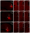
The principal cellular elements of the mammalian SVZ evolved prior to the appearance of modern day mammals and were likely present in the ancestor to both mammal and sauria. (A–G) Position of Tbr2+ neural progenitor cells in the developing cortex of mammalian and non-mammalian species. Intermediate progenitor cells labeled with Tbr2 (red) are located within the cortical proliferative zones in a distinct fashion in each species. All cell nuclei are labeled with DAPI (blue). (A) Low power image of the primary sensory macaque cerebral cortex at gestation day 80. (B) Higher magnification image from panel (A). The outer SVZ (oSVZ) is substantially thicker, and the total number of Tbr2+ cells is greater in rhesus macaques neocortex compared to non-primate species. Images in panels B–G are shown at the same scale. (C) P2 developing ferret neocortex. (D) E20 developing rat neocortex. The thickness of the SVZ in the lissencephalic rat is similar to that present in the gyrencephalic ferret, but significantly thinner than in macaque. The number of Tbr2+ cells in a given section of brain tissue from the developing ferret (C) and rat (D) neocortex is similar. (E) E8 chick pallium. Tbr2+ cells are positioned within a robust SVZ. (F) In stage 20 turtle pallium Tbr2+ cells are dispersed throughout the VZ in the developing dorsal cortex. However, in the dorsal ventricular ridge (DVR) Tbr2+ cells are organized into a tight subventricular band. (G) Stage 11 lizard pallium. Tbr2+ cells in lizard are dispersed throughout the VZ. (H) Cladogram showing the relationship between mammals, turtles, birds, crocodiles, lizards, and snakes. Based on recent genetic analysis [49], turtles and birds have been placed in the recently proposed clade archelosauria, while lizards and other reptiles, such as snakes, are in the superorder lepidosauria. Mammals and birds possess an SVZ based on the distribution of Tbr2+ cells (red asterisks). Tbr2+ cells are present in both the dorsal cortex and dorsal ventricular ridge (DVR) of developing turtles but only the DVR shows evidence of an SVZ (red/black asterisk). Tbr2+ cells are also present in the developing lizard and crocodile forebrain, but there is no evidence of abventricular divisions or an anatomically defined SVZ (black asterisk). Interestingly, the developing snake cortex presented with a different structure than that of reptiles and crocodiles, being more similar to that of mice where Tbr2+ cells were organized into a tight SVZ band that was superficial to the VZ. The presence of Tbr2+ cells and an SVZ in mammals and both reptilian clades, archelosauria and lepidosauria, supports the concept that the common ancestor for mammals and reptiles possessed Tbr2+ cells and an SVZ. Scale bar = 250 μm, and applies to B–G.
Figure 2
Figure 2
Mammalian, but not avian, forebrain progenitor cells labelled through Emx2 promoter sequence show delayed neurogenesis (García-Moreno and Molnár, 2015). (A) Top row - Examples of the distribution of the cortical neurons in mouse E14, 2 days after electroporation with general and Emx2 promoter construct at E12. Bottom row - Chick cases at E6, 2 days after electroporation at E4. Left column - Labelled with EGFP (yellow), early progenitors of the forebrain generated neurons that migrated to the postmitotic areas (CP: cortical plate; MZ: mantle zone). Right column - Also labelled with EGFP, early progenitors selected by their expression of Emx2 did not generate neurons in mouse (observe the absence of yellow cells in CP) but were neurogenic in chick (green cells were present in MZ). (B) Schematic summary of the differences found in the development of the dorsal forebrain in mouse and chick. In mouse cortex there are progenitors that contribute to all layers and an early delayed progenitor subtype that mostly contributes to upper layers. In chick there is no such distinction between the differentially labelled progenitors.
Figure 3
Figure 3
Evolution of cortical circuits in amniotes. (A) The reptilian three-layered dorsal cortex and the mammalian six-layered neocortex develop from homologous pallial regions and display analogous properties at the functional level (blue and red: glutamatergic and GABAergic neurons). However, it is not yet clear how reptilian and mammalian cortices compare at the circuit level. Do they share evolutionarily conserved neuronal types? Do these cell types participate in common microcircuit motifs? (B) Tosches and colleagues are profiling reptilian cortical neurons using single cell transcriptomics. Microfluidics devices are used to capture individual cells from dissociated tissue and to obtain single-cell cDNAs for deep sequencing. Multivariate statistical approaches, like PCA and clustering, are used to identify molecularly defined neuronal types. These data contribute to our understanding of cortical evolution, and provide the foundation for building new molecular tools to study circuit function in reptiles.
Figure 4
Figure 4
The arcopallium projects domain-specifically onto the nucleus accumbens (also representative of other ventrobasal forebrain projections). (A–A‴) D594+ axons traced anterogradely from the amygdalopiriform area (APir) of the arcopallium, terminating in both the rostral, intermediate and caudal parts of the nucleus accumbens (Ac) in great density. Representative insets demonstrate varicose axons under high magnification. (B–B‴) Injection placed into the dorsal arcopallium (ADo) labeled fewer terminals throughout the Ac. Representative insets demonstrate varicose axons under high magnification, though such axons were rather sporadic in the rostralmost Ac. (C–C‴) The medial, hilar division of the arcopallium (AHil) gave rise to axons that terminated in the caudal and intermediate, but not the rostral, part of the Ac. Note the low density of terminal axons even in the intermediate and caudal parts of the Ac. Representative insets demonstrate varicose axons under high magnification. Here, due to an overall scarcity of varicose fibers, no such element could be indicated in the rostralmost Ac. (A, B, C) Appropriate symbols indicate the injection sites. Due to low intensity of section images (optimized for the fluorescent signal of tracer deposit), the outlines of sections are indicated by doted lines. (A–C‴) Cranio-caudal levels of coronal sections are indicated as distance in millimeters AP according to Kuenzel and Masson (1988). Abbreviations: D594 Alexa Fluor® 594 conjugated high-molecular-weight (10kDa) dextran, dors dorsal, med medial. Scale bars: 1mm (A, B, C), 70μm (A′-A‴, B′-B‴, C′-C‴), 2μm (insets in A′-A‴, B′-B‴, C′-C‴). Reproduced with author’s modification from Hanics et al, 2016 (Brain Structure and Function).
Figure 5
Figure 5
Photomicrograph of the hippocampal formation in the 500 g brain of the harbour porpoise (Phocoena phocoena) showing the small size and loose architectural organization of this structure in the cetacean brain. The hippocampal formation of cetaceans is around 5 times smaller than one would expect for a mammal with the brain size of cetaceans (Patzke et al., 2015), and with either a very small or absent prefrontal cortex (Manger, 2006) it is unclear whether the cognitive functions associated with the hippocampus and prefrontal cortex in other mammals, such as the encoding, retrieval, long term storage and contextualization of memories, will be functionally relevant in the cetaceans. Scale bar = 1 mm. CA–cornu ammonis region; DG–dentate gyrus.

References

    1. Martinez-Cerdeno V, Cunningham CL, Camacho J, Keiter JA, Ariza J, Lovern M, Noctor SC. Evolutionary origin of Tbr2-expressing precursor cells and the subventricular zone in the developing cortex. The Journal of comparative neurology. 2016;524(3):Spc1. doi: 10.1002/cne.23949. - DOI - PMC - PubMed
    1. Martinez-Cerdeno V, Cunningham CL, Camacho J, Antczak JL, Prakash AN, Cziep ME, Walker AI, Noctor SC. Comparative analysis of the subventricular zone in rat, ferret and macaque: evidence for an outer subventricular zone in rodents. PloS one. 2012;7(1):e30178. doi: 10.1371/journal.pone.0030178. Epub 2012/01/25. - DOI - PMC - PubMed
    1. Martinez-Cerdeno V, Noctor SC, Kriegstein AR. The role of intermediate progenitor cells in the evolutionary expansion of the cerebral cortex. Cereb Cortex. 2006;16(Suppl 1):i152–i61. - PubMed
    1. Kriegstein A, Noctor S, Martinez-Cerdeno V. Patterns of neural stem and progenitor cell division may underlie evolutionary cortical expansion. Nature reviews Neuroscience. 2006;7(11):883–90. doi: 10.1038/nrn2008. Epub 2006/10/13 nrn2008 [pii] - DOI - PubMed
    1. Smart IH, Dehay C, Giroud P, Berland M, Kennedy H. Unique morphological features of the proliferative zones and postmitotic compartments of the neural epithelium giving rise to striate and extrastriate cortex in the monkey. Cereb Cortex. 2002;12(1):37–53. - PMC - PubMed

Publication types