Review
. 2017 Jan:34:E011.
doi: 10.1017/S0952523817000050.

The mouse pulvinar nucleus: Organization of the tectorecipient zones

Affiliations
Review

The mouse pulvinar nucleus: Organization of the tectorecipient zones

N A Zhou et al. Vis Neurosci. 2017 Jan.

Abstract

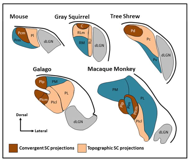
Comparative studies have greatly contributed to our understanding of the organization and function of visual pathways of the brain, including that of humans. This comparative approach is a particularly useful tactic for studying the pulvinar nucleus, an enigmatic structure which comprises the largest territory of the human thalamus. This review focuses on the regions of the mouse pulvinar that receive input from the superior colliculus, and highlights similarities of the tectorecipient pulvinar identified across species. Open questions are discussed, as well as the potential contributions of the mouse model for endeavors to elucidate the function of the pulvinar nucleus.

Keywords: Superior colliculus; Synapse; Thalamus; Visual.


Figures

Figure 1
Figure 1. The pulvinar nucleus contains two tectorecipient zones
Schematic illustrations indicate regions of the pulvinar nucleus in the mouse, squirrel, tree shrew, galago and macaque monkey that have been shown to receive dense convergent input (brown) or less dense topographic projections (peach) from the superior colliculus. The non-tectorecipient zones of the pulvinar are indicated in blue, and the location of the dorsal lateral geniculate nucleus (dLGN, gray) is indicated for reference. Illustrations are not to scale (adapted from Baldwin et al., 2013, 2011; Chomsung et al., 2008; Day-Brown et al., 2016; Stepniewska et al., 2000). Subdivisions for Mouse: Pcm, caudal medial pulvinar, Pl, lateral pulvinar, Prm, rostral medial pulvinar, Squirrel: C, caudal pulvinar, RL, rostral lateral pulvinar, RLm, medial rostral lateral pulvinar, RLl, lateral rostral lateral pulvinar, RM, rostral medial pulvinar, Tree shrew: Pc, central pulvinar, Pd, dorsal pulvinar, Pv, ventral pulvinar, Galago and Macaque: PIcm, central medial inferior pulvinar, PIcl, central lateral inferior pulvinar, PIp, posterior inferior pulvinar, PIpl, posterior lateral inferior pulvinar, PL, lateral pulvinar, PM, medial pulvinar, Macaque: PIm, medial inferior pulvinar.
Figure 2
Figure 2. Widefield vertical (WFV) cells project to the ipsilateral and contralateral pulvinar
Panel A illustrates an injection of a retrogradely transported virus (MIT viral vector core: hEF1α-EYFP-IRES-cre) in the pulvinar (PUL) of a wild type mouse that induced the expression of yellow fluorescent protein (YFP, green) in WFV cells of the superior colliculus (SC). Cells labeled by this injection are illustrated in panel B in a contralateral SC section that was stained with an antibody against calretinin (purple), which delineates the stratum griseum superficiale (SGS). The WFV tectopulvinar cells are located in the stratum opticum (SO) and lower SGS and extend dendrites to the surface of the SC, where they end in complex dendritic tufts (panel C). Panels D and E illustrate WFV cells labeled by injections of retrogradely transported cre-dependent viruses (MIT-viral vector core: hEF1α-LS1L-mCherry and hEF1α-LS1L-EYFP) in the left and right pulvinar of a substance P-cre mouse (Jackson Labs stock number 021877) to induce the expression of either YFP (green, left pulvinar injection) or mCherry (purple, right pulvinar injection) in cre-expressing neurons. Many WFV cells expressed both YFP and mCherry (white), demonstrating that a subpopulation of WFV cells bilaterally innervate the pulvinar, and that WFV cells express substance P. Scale bars: A and B = 100 μm, C = 10 μm, D = 50 μm and also applies to E. dLGN, dorsal lateral geniculate nucleus, PT, pretectum, OT, optic tract. Virus injection methods as in Bickford et al. (2015).
Figure 3
Figure 3. Caudal medial pulvinar (Pcm) cells express calretinin (CR) and neurokinin 1 (NK1) and align with bilateral superior colliculus (SC) projections
Confocal images illustrate ipsilateral (A, C, green) and contralateral (D, F, green) projections to the pulvinar that were labeled by a unilateral virus injection in the SC. These sections were also stained with antibodies against CR (B, E, purple) to define the Pcm (which contains CR) and the lateral pulvinar (Pl, which does not contain CR). Adjacent sections (C, F) stained for CR (purple) and NK1 (green) illustrate that CR-positive Pcm cells express NK1. This expression pattern is shown at higher magnification in half micron optical sections in panels G (CR, purple), H (NK1, green) and I (CR, purple, and NK1, green, asterisks indicate cells labeled with both antibodies). Scale in A = 50 μm and applies to A–F. Scale in G = 10 μm and applies to G–I. dLGN, dorsal lateral geniculate nucleus, OT, optic tract, PT, pretectum. Methods as in Bickford et al. (2015) and Masterson et al. (2010).
Figure 4
Figure 4. The Pcm contains a dense population of terminals that contain substance P
A–C) Caudal to rostral sections stained with an antibody against substance P (visualized with a diaminobenzidine reaction). Staining is densest in the caudal and medial pulvinar (Pcm). Little staining is observed in the lateral pulvinar (Pl). Scale = 100 μm and applies to all panels. dLGN, dorsal lateral geniculate nucleus, MGN, medial geniculate nucleus, OT, optic tract, PT, pretectum. Methods as in Masterson et al. (2010).
Figure 5
Figure 5. Ultrastructure of cortical and tectal terminals in the mouse pulvinar
Terminals labeled by the anterograde transport of biotinylated dextran amine injected in V1 (A), superior colliculus (B) or the posterior/postrhinal cortex (C) contact (white arrows) the proximal (A, B) and distal (C) dendrites of pulvinar neurons (green overlay). Sections were additionally stained with gold particles to reveal the distribution of GABA. This indentifies two types of GABAergic terminals (purple overlay) in the mouse pulvinar: F2 profiles (B) contain a low density of vesicles and F1 profiles (C) contain a high density of vesicles. Scale = 600 nm and applies to all panels. Methods as in Li et al. (2003c).
Figure 6
Figure 6. Tectopulvinar and corticopulvinar terminals overlap in the caudal medial (Pcm) and lateral (Pl) subdivisions of the mouse pulvinar
This overlap is demonstrated via dual virus injections in the superior colliculus (SC) and lateral extrastriate cortex (LES, first 2 columns, A–D and E–H), or SC and V1 (last column, I–L). The Pcm and Pl subdivisions are defined using immunocytochemical staining for calretinin (CR, blue, first row, A, E, I). Virus injections were placed in the SC to induce the expression of yellow fluorescent protein (green, panels B, F, J), and in the cortex (V1 or LES) to induce the expression of TdTomato (red, panels C, G, K), Overlap of the CR and virus labeling patterns (panels D, H, L) show that the Pcm is innervated by the SC and LES, while the Pl is innervated by the SC, V1 and LES (panels D, H, L). Scale bar in D = 100 μm and applies to all panels. dLGN, dorsal lateral geniculate nucleus, PT, pretectum. Methods as in Bickford et al. (2015) and Jurgens et al. (2012).
Figure 7
Figure 7. Potential input integration in the mouse pulvinar
Terminals labeled by a virus injection in V1 (green, A, B) and the ipsilateral SC (purple, B) overlap in the Pl. C) Two biocytin-filled pulvinar neurons (green) and surrounding tectopulvinar terminals (purple, labeled by a virus injection in the ipsilateral SC). The dendrites of the pulvinar neurons extend across subdivisions. D) Biocytin-filled pulvinar interneurons (purple) identified in a mouse line (Jackson Laboratories stock number 007677) that expresses green fluorescent protein in GABAergic neurons (green) extend dendrites across subdivisions. Scale bars = 20 μm. Methods as in Bickford et al., (2015).
Figure 8
Figure 8. The mouse pulvinar projects to the cortex, striatum and amygdala
Injections of biotinylated dextran amine in the mouse pulvinar (A) label terminals in V1 (B) and extrastriate cortex regions including the posterior medial area (PM, panel B) and the lateral medial area (LM, panel D). Cells in the superior colliculus (C) and LM (D) are also labeled by retrograde transport. E) The pulvinar also projects to the caudate and putamen (CPu) and lateral amygdala (LA). Scale = 200 μm and applies to all panels. Methods as in Chomsung et al. (2010).
Figure 9
Figure 9. The tectorecipient mouse pulvinar forms interconnected loops with the cortex, striatum and amygdala
The schematic diagrams illustrate the main connections of the tectorecipient subdivisons of the mouse pulvinar. The caudal medial pulvinar (Pcm, red) receives bilateral input from widefield vertical (WFV) cells of the superior colliculus, and is reciprocally connected to the posterior (P) and postrhinal (POR) regions of the cortex, where it innervates layers I and IV–VI. Both the Pcm and P/POR project to the caudal caudate/putamen (CPu) and lateral amygdala (LA). The lateral pulvinar (Pl, blue) receives ipsilateral input from WFV cells, and is reciprocally connected to V1 and the lateral medial (LM) and lateral intermediate (LI) regions of the cortex. Within V1, the Pl projects to layers I and Va. Within LM and LI, the Pl projects to layer I and IV. The Pl, LM and LI project to the middle regions of the CPu.

Similar articles

Cited by

References

    1. Abramson BP, Chalupa LM. The laminar distribution of cortical connections with the tecto- and cortico-recipient zones in the cat’s lateral posterior nucleus. Neuroscience. 1985;15:81–95. - PubMed
    1. Abramson BP, Chalupa LM. Multiple pathways from the superior colliculus to the extrageniculate visual thalamus of the cat. J Comp Neurol. 1988;271:397–418. - PubMed
    1. Allen AE, Procyk CA, Howarth M, Walmsley L, Brown TM. Visual input to the mouse lateral posterior and posterior thalamic nuclei: photoreceptive origins and retinotopic order. J Physiol. 2016;594:1911–1929. - PMC - PubMed
    1. Baldwin MKL, Wong P, Reed JL, Kaas JH. Superior colliculus connections with visual thalamus in gray squirrels (Sciurus carolinensis): evidence for four subdivisions within the pulvinar complex. J Comp Neurol. 2011;519:1071–1094. - PMC - PubMed
    1. Baldwin MKL, Balaram P, Kaas JH. Projections of the superior colliculus to the pulvinar in prosimian galagos (Otolemur garnettii) and VGLUT2 staining of the visual pulvinar. J Comp Neurol. 2013;521:1664–1682. - PMC - PubMed

Publication types

LinkOut - more resources