Review
. 2017 Jan:34:E013.
doi: 10.1017/S0952523817000104.

An evolving view of retinogeniculate transmission

Affiliations
Review

An evolving view of retinogeniculate transmission

Elizabeth Y Litvina et al. Vis Neurosci. 2017 Jan.

Abstract

The thalamocortical (TC) relay neuron of the dorsoLateral Geniculate Nucleus (dLGN) has borne its imprecise label for many decades in spite of strong evidence that its role in visual processing transcends the implied simplicity of the term "relay". The retinogeniculate synapse is the site of communication between a retinal ganglion cell and a TC neuron of the dLGN. Activation of retinal fibers in the optic tract causes reliable, rapid, and robust postsynaptic potentials that drive postsynaptics spikes in a TC neuron. Cortical and subcortical modulatory systems have been known for decades to regulate retinogeniculate transmission. The dynamic properties that the retinogeniculate synapse itself exhibits during and after developmental refinement further enrich the role of the dLGN in the transmission of the retinal signal. Here we consider the structural and functional substrates for retinogeniculate synaptic transmission and plasticity, and reflect on how the complexity of the retinogeniculate synapse imparts a novel dynamic and influential capacity to subcortical processing of visual information.

Keywords: Developmental refinement; Retinogeniculate synapse; Short-term plasticity; Synaptic transmission; Visual circuit.


Figures

Fig. 1.
Fig. 1.
Synaptic structure shapes retinogeniculate transmission. (A) Tracing of an HRP-filled X-RGC arbor in the cat dLGN shows the location and morphology of a single branch (red box) of the X-RGC arbor used for EM reconstruction. This branch of the axon contacts 4 TC neurons out of 40 available neurons in the territory of the arbor. The remainder of the axon was not reconstructed, and likely contacts several other TC neurons. Bottom inset shows the location of the axonal arbor in the context of the cat LGN. Figure modified from Hamos et al. (1987). Unmarked scale bar = 100 μm. (B, C) Reconstructed arbors of single RGC axons showing distribution of presynaptic boutons into dense clusters in the LGN of (B) an adult cat and (C) a p20 mouse. Note the clustering of boutons along the arbor. Image in B is modified from Robson et al. (1993), showing a segment of a RGC axon; Image in C is from Hong et al. (2014), showing a BD-RGC axon. Scales bars are 100 μm. (D) A 3D reconstruction of a TC neuron dendrite and sites of contact between two neighboring RGC boutons from Budisantoso et al. (2012). In the top image, the dendrite and its appendages are depicted in blue, whereas pink and red sites label the postsynaptic densities of the two axons. In the bottom image, the structure of the terminals of two axons has been added. Spillover can occur between these two nearby terminals. (E) Evidence of spillover-mediated responses to the stimulation of a single RGC axon before eye opening. Two different synaptic responses were observed in response to single retinal fiber stimulation. Shown are recordings from TC neurons in whole cell voltage clamp at −70 mV in a dLGN slice in the presence of the NMDAR blocker, 20 μM CPP. On the left is an example of a retinogeniculate AMPAR EPSC with characteristic rapid rise time and decay kinetics (black trace). On the right is an atypical AMPAR EPSC response notable for significantly slower rise time and decay kinetics (black trace). The two types of EPSCs differ in their sensitivity to the low-affinity AMPAR antagonist, γ−DGG. Low affinity antagonists can be used to assess the relative concentration of glutamate in the synaptic cleft (Clements et al., 1992; Diamond & Jahr, 1997). As γ−DGG competes with glutamate for binding to AMPAR, its efficacy of inhibition decreases with increasing glutamate concentration. γ−DGG has only a small effect on the amplitude of the fast EPSC, but dramatically reduces the amplitude of the slow EPSC (overlaid gray traces), consistent with lower peak glutamate concentration in the synaptic cleft of the slow EPSC. Because the EPSCs are evoked by minimal stimulation, the rapid EPSC represents a direct input from a single RGC axon that forms a direct synapse onto the voltage-clamped relay neuron, whereas the slow EPSC corresponds to the activation of a RGC axon that does not directly synapse onto the voltage-clamped neuron. Modified from Hauser et al. (2014). All figures reprinted with permission.
Fig. 2.
Fig. 2.
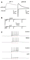
Contributions of retinogeniculate short-term plasticity. (A) Representative traces of AMPAR and NMDAR mediated currents recorded before eye opening (left) and in a mature mouse (right) in response to the stimulation of the optic tract. Whole-cell voltage clamp recordings were performed with bicuculline to block GABAA−receptor mediated currents. At −70 mV holding potential, AMPARs mediate the fast activating and decaying current. AMPAR and NMDAR currents both contribute to the EPSCs recorded at +40 mV with AMPARs contributing to the rapid rise and the NMDAR currents contributing to the slow decay of the EPSC. The average amplitude of AMPAR currents increases over development. (B) 5-CT-mediated activation of serotonin receptors alters retinogeniculate short-term plasticity. Experiments were performed in retinogeniculate slices from mature mice. Top and bottom traces overlay pairs of retinogeniculate EPSCs evoked with varying ISI before (top) and after (bottom) the application of 5-CT to active 5HT-1 receptors expressed in presynaptic retinogeniculate boutons. Application of 5-CT reduces the amplitude of the first EPSC and relieves short-term depression, increasing the amplitude of the second EPSC preferentially at short interstimulus interval. (C) Physiologically relevant stimulation frequencies preferentially diminish the contribution of AMPARs to relay neuron firing. Current clamp recordings of action potential firing in response to trains of optic tract stimulation in the presence of AMPAR (NBQX) or NMDAR (CPP) antagonists. Holding potential −50 mV. Blockade of AMPARs alters the latency to first spike but only minimally reduces the overall number of spikes. In contrast, blockade of NMDARs abolished EPSC summation toward action potential firing; only the first stimulus evokes an action potential, reflecting the contribution of AMPARs that rapidly desensitize after the first pulse. Therefore, NMDAR currents can sustain action potential generation without AMPAR contribution. Adapted from (A) Chen and Regehr (2000), B) Liu and Chen (2008) and (C) Augustinaite and Heggelund (2007). All figures reprinted with permission.
Fig. 3.
Fig. 3.
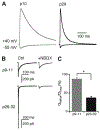
Contribution of NMDAR-currents to retinogeniculate transmission over development. (A) NMDAR EPSCs recorded in the presence of the AMPAR blocker, NBQX, at +40 and −55 mV holding potentials in a p10 (left) and a p29 (right) retinogeniculate slice. Normalized traces are shown. Note the acceleration in NMDAR current decay time over development. (B) Example EPSCs recorded in young (top) and mature (bottom) TC neuron in slice before (left) and during (right) the application of NBQX. Holding potential, −55 mV. (C) NMDAR currents contribute more to the total retinogeniculate charge transfer at p9–11 than p26–32; however, even at the mature synapse, NMDARs contribute nearly half of the total charge transfer. Figure adapted from Liu and Chen (2008). All figures reprinted with permission.
Fig. 4.
Fig. 4.
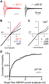
Substrates for retinogeniculate plasticity. (A) Overlaid AMPAR current traces recorded from different holding potentials to assess the current voltage (IV) relationship. Currents in the presence of CPP to block NMDAR currents and with spermine in the internal solution to examine the degree of IV rectification. Calcium-permeable AMPARs exhibit a rectifying IV relationship. Traces were recorded at 20 mV increments from −60 to +60 mV holding potentials. Left-example obtained before eye opening; right, example from a mature slice. From Hauser et al. (2014). (B) Change in the average AMPAR EPSC IV relationship over development. Rectification of IV currents increases significantly from p9–11 to maturity, indicating a gradual increase in the contribution of CP-AMPARs to AMPAR-mediated currents. Modified from Hauser et al. (2014). Red: p9–11; blue: p15–16; black: p27–32. (C) Changes in AMPAR subunit composition in response to visual experience. The effect of visual deprivation from p20 (late-dark rear, LDR) or dark rearing from birth (chronic dark reared, CDR) on the AMPAR EPSC IV relationship. Rectification of AMPAR currents is reduced in LDR but not in chronically dark reared (CDR) mice when compared to normally reared mice (light rear, LR) mice. P = 0.03. Recordings performed at p27–32. Modified from Louros et al. (2014). (D) Comparison of the distribution of amplitudes of single fiber RGC inputs in juvenile (p27–34) and adult (p60+) mice show the persistence of weak (small-amplitude) inputs with age. Modified from Hong et al. (2014). All figures reprinted with permission.

Similar articles

Cited by

References

    1. Ackman JB, Burbridge TJ & Crair MC (2012). Retinal waves coordinate patterned activity throughout the developing visual system. Nature 490, 219–225. - PMC - PubMed
    1. Aguila J, Cudeiro FJ & Rivadulla C (2017). Suppression of V1 feedback produces a shift in the topographic representation of receptive fields of LGN cells by unmasking latent retinal drives. Cerebral Cortex 27, 3331–3345. - PubMed
    1. Akerman CJ, Smyth D & Thompson ID (2002). Visual experience before eye-opening and the development of the retinogeniculate pathway. Neuron 36, 869–879. - PubMed
    1. Albrecht D, Davidowa H & Gabriel H (1990). Conditioning-related changes of unit activity in the dorsal lateral geniculate nucleus of urethane-anaesthetized rats. Brain Research Bulletin 25, 55–63. - PubMed
    1. Alitto HJ, Moore BD, Rathbun DL & Martin Usrey W (2011). A comparison of visual responses in the lateral geniculate nucleus of alert and anaesthetized macaque monkeys. The Journal of Physiology 589, 87–99. - PMC - PubMed

Publication types

MeSH terms

LinkOut - more resources