Review
. 2018 Mar;32(2):96-105.
doi: 10.1016/j.blre.2017.09.002. Epub 2017 Sep 18.

Erythroleukemia-historical perspectives and recent advances in diagnosis and management

Affiliations
Review

Erythroleukemia-historical perspectives and recent advances in diagnosis and management

Prajwal Boddu et al. Blood Rev. 2018 Mar.

Abstract

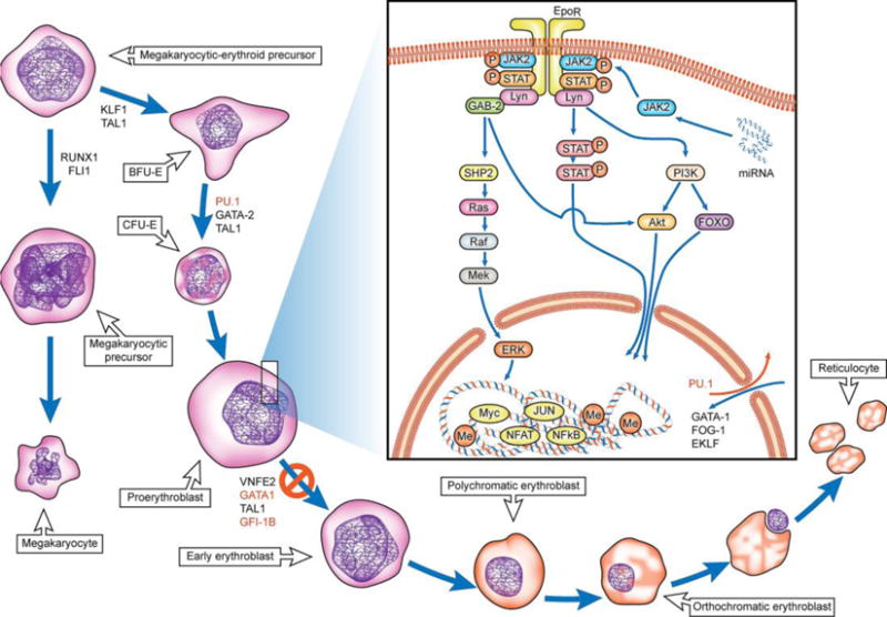
Acute erythroleukemia is a rare form of acute myeloid leukemia recognized by its distinct phenotypic attribute of erythroblastic proliferation. After a century of its descriptive history, many diagnostic, prognostic, and therapeutic implications relating to this unique leukemia subset remain uncertain. The rarity of the disease and the simultaneous involvement of its associated myeloid compartment have complicated in vitro studies of human erythroleukemia cell lines. Although murine and cell line erythroleukemia models have provided valuable insights into pathophysiology, translation of these concepts into treatment are not forthcoming. Integration of knowledge gained through a careful study of these models with more recent data emerging from molecular characterization will help elucidate key mechanistic pathways and provide a much needed framework that accounts for erythroid lineage-specific attributes. In this article, we discuss the evolving diagnostic concept of erythroleukemia, translational aspects of its pathophysiology, and promising therapeutic targets through an appraisal of the current literature.

Keywords: Acute erythroleukemia – M6a subtype; Acute erythroleukemia – M6b subtype; Bromodomain protein; Erythroblasts; GATA1 protein; MicroRNA; PU.1 protein; Pure erythroid leukemia; TP53.


Conflict of interest statement

Conflict of Interest

The authors declare no conflicts of interest in the publication of this paper.

Figures

Figure 1
Figure 1
Modern diagnostic approach to acute erythroid leukemia. M6 leukemia subtype is highlighted in yellow. Pure erythroid leukemia remains only the true form of erythroleukemia based on the updated WHO 2016 classification. Abbreviations used: MRC-myelodysplasia related changes, NOS- not otherwise specified.
Figure 2
Figure 2
A, Bone marrow core biopsy of pure erythroid leukemia often shows sheets of immature erythroid precursors replacing marrow cellular spaces. Cells are large with round to irregular nuclei, pale chromatin and frequent one to several distinct nucleoli. Background trilineage hematopoiesis is significantly decreased to absent. B, CD71 immunostain highlights leukemic cells with strong membranous staining pattern. C, on bone marrow smears, leukemic cells have dispersed chromatin, deep basophilic cytoplasm and cytoplasmic vacuoles. Some also have cytoplasmic blebs. Background erythroid dysplasia is present in this case. D. Coarsely granular pattern by PAS stain.
Figure 3
Figure 3
Model for erythroid differentiation and pathways implicated in human/murine erythroleukemia. Scheme outlines the stages of differentiation and maturation from the common megakaryocytic-erythroid precursor stage (70). The transcription factors, signaling proteins and miRNAs aberrantly expressed in human erythroleukemia models are highlighted in red (87). There exists a differentiation blockade at the proerythroblast stage preventing further differentiation to more mature blasts. The underlying mechanisms responsible for the blockade are an active area of investigation.
Figure 4
Figure 4
Proposed potential therapeutic targets in Erythroleukemia. A wide range of signaling pathway mutations (JAK2, NTRK1, ALK), epigenetic alterations (Hypomethylating agents, LSD-1 inhibitors, bromodomain inhibitors), and microRNA dysregulation (microRNA therapies) present with multiple options for therapeutic targeting. Abbreviations used: BRD-bromodomain reading proteins, miR-microRNA, EpoR-Erythropoietin receptor
Figure 5
Figure 5
A proposed two-step model of erythroleukemia. Epigenetic and micro-RNA dysregulation, and transcription factor level alterations lead to differentiation arrest at the pro-erythroblast stage. Arrested pro-erythroblasts are hyper-proliferative and accumulate secondary mutations, including TP53, leading to leukemic transformation of arrested pro-erythroblasts. Continued maintenance and proliferation of leukemic blasts is facilitated by pre-existing genetic/epi-genetic alterations in the already transformed blasts.

Similar articles

Cited by

References

    1. Schwartz SO, Critchlow J. Erythremic myelosis (DI Guglielmo’s disease); critical review with report of four cases, and comments on erythroleukemia. Blood. 1952;7(8):765–93. - PubMed
    1. Dameshek W, Baldini M. The Di Guglielmo syndrome. Blood. 1958;13:192–4. - PubMed
    1. Bain BJ. Di Guglielmo and his syndromes. Br J Haematol. 2003;120:939–43. - PubMed
    1. Dameshek W. Some speculations on the myeloproliferative syndromes. Blood. 1951;6(4):372–5. - PubMed
    1. Dameshek W. The DiGuglielmo syndrome revisited. Blood. 1969;34(5):567–72. - PubMed

Publication types