. 2017 Oct 5;101(4):552-563.
doi: 10.1016/j.ajhg.2017.08.017. Epub 2017 Sep 28.

Hypomorphic Recessive Variants in SUFU Impair the Sonic Hedgehog Pathway and Cause Joubert Syndrome with Cranio-facial and Skeletal Defects

Affiliations

Hypomorphic Recessive Variants in SUFU Impair the Sonic Hedgehog Pathway and Cause Joubert Syndrome with Cranio-facial and Skeletal Defects

Roberta De Mori et al. Am J Hum Genet. .

Abstract

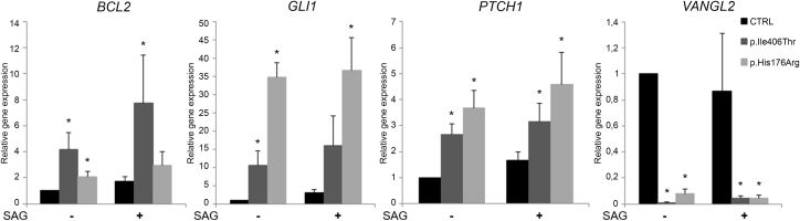
The Sonic Hedgehog (SHH) pathway is a key signaling pathway orchestrating embryonic development, mainly of the CNS and limbs. In vertebrates, SHH signaling is mediated by the primary cilium, and genetic defects affecting either SHH pathway members or ciliary proteins cause a spectrum of developmental disorders. SUFU is the main negative regulator of the SHH pathway and is essential during development. Indeed, Sufu knock-out is lethal in mice, and recessive pathogenic variants of this gene have never been reported in humans. Through whole-exome sequencing in subjects with Joubert syndrome, we identified four children from two unrelated families carrying homozygous missense variants in SUFU. The children presented congenital ataxia and cerebellar vermis hypoplasia with elongated superior cerebellar peduncles (mild "molar tooth sign"), typical cranio-facial dysmorphisms (hypertelorism, depressed nasal bridge, frontal bossing), and postaxial polydactyly. Two siblings also showed polymicrogyria. Molecular dynamics simulation predicted random movements of the mutated residues, with loss of the native enveloping movement of the binding site around its ligand GLI3. Functional studies on cellular models and fibroblasts showed that both variants significantly reduced SUFU stability and its capacity to bind GLI3 and promote its cleavage into the repressor form GLI3R. In turn, this impaired SUFU-mediated repression of the SHH pathway, as shown by altered expression levels of several target genes. We demonstrate that germline hypomorphic variants of SUFU cause deregulation of SHH signaling, resulting in recessive developmental defects of the CNS and limbs which share features with both SHH-related disorders and ciliopathies.

Keywords: GLI3; Joubert syndrome; SUFU; Sonic Hedgehog; ciliopathies; congenital ataxia; developmental defects; hypomorphic variants; molar tooth sign; polymicrogyria.


Figures

Figure 1
Figure 1
Clinical and Neuroimaging Features of Subjects Harboring SUFU Homozygous Missense Variants (A–D) Photographs of the four children from families COR369 (A, B) and MTI-2023 (C, D). Note hypertelorism, frontal bossing, and broad, depressed nasal bridge. (E and F) Bilateral hand post-axial polydactyly (E) and plantar dyskeratotic pits (F) in the male sibling from family COR369. (G) Right hand post-axial polydactyly in the male sibling from family MTI-2023. (H–O) Brain MRI of affected girl (H–K) and boy (L–O) from family COR369. Note mild vermis hypoplasia (H, L) and elongated, thickened, and horizontally orientated superior cerebellar peduncles consistent with mild MTS (I, M), associated with bilateral polymicrogyria mainly involving the right perisylvian regions (arrows in J, K, N, O). Also note the megacisterna magna in the sagittal image of the girl (H). (P–S) Brain MRI in the proband from family MTI-2023. Note vermis hypoplasia, megacisterna magna (P, Q), dysplastic foliar abnormalities (curved arrows in R), deep interpeduncular fossa, and thickened horizontally orientated superior cerebellar peduncles (mild MTS, Q). In all subjects, mild lateral ventricular enlargement and shrunken white matter in the supratentorial posterior regions are visible (K, N, O, S). MRI of the fourth child were not available for publication.
Figure 2
Figure 2
Analysis of MD Simulations of WT and Mutant SUFU Proteins (A) 3D structure of SUFU-GLI complex. The N-terminal (28–262), C-terminal (266–285; 345–480), and linked (263–265) domains are colored green, yellow, and gray, respectively, while the GLI3 protein is colored in red. p.Ile406Thr is highlighted in blue and p.His176Arg in orange. (B) RMSD of all heavy atoms of SUFU. (C) RMSF of SUFU residues during simulations. (D) Covariance matrix for SUFU WT (top), SUFUp.Ile406Thr (center), and SUFUp.His176Arg (bottom). Perfect direct or inverse correlations are highlighted in red or violet, respectively. (E) Distances between amino acids forming the binding site for GLI3. (F) H-bonds established during simulation between the residues forming the ligand binding pocket and GLI3. In (B)–(F) graphs, SUFU WT is colored in black, SUFUp.Ile406Thr in red, and SUFUp.His176Arg in green.
Figure 3
Figure 3
SUFU Missense Variants Reduce Protein Stability (A) IMCD3 cells transfected with HA-tagged SUFU WT, SUFUp.Ile406Thr, and SUFUp.His176Arg for 24, 48, and 72 hr show a more rapid decrease of mutant SUFU protein levels compared to WT (quantified by densitometry in the corresponding graphs). (B) Control and mutant fibroblasts were treated with cycloheximide for 24 hr. Mutant cells show significantly lower levels of SUFU even in basal conditions, with a further more marked decrease after treatment. Results are representative of at least three independent experiments. Graphs represent means ± standard errors. p < 0.05.
Figure 4
Figure 4
SUFU Missense Variants Impair Binding to GLI3 (A) Co-immunoprecipitation experiments in IMCD3 cells transfected with either HA-tagged SUFU WT, SUFUp.Ile406Thr, or SUFUp.His176Arg, showing reduced binding of GLI3 to mutant SUFU. (B) GLI3 full length and GLI3R levels in mutant fibroblasts are significantly lower than in controls in basal conditions, with a further decrease after 24 hr SAG stimulation. Results are representative of at least four independent experiments. Graphs represent means ± standard errors. p < 0.05.
Figure 5
Figure 5
The SHH Pathway Is Deregulated in Mutant Fibroblasts Histograms show the expression levels of four target genes that in basal conditions are either significantly overexpressed (BCL2, GLI1, and PTCH1) or under-expressed (VANGL2) in fibroblasts from the two probands compared to control subjects. Results are representative of three independent experiments. Graphs represent means ± standard errors. p < 0.05.

References

    1. Gorojankina T. Hedgehog signaling pathway: a novel model and molecular mechanisms of signal transduction. Cell. Mol. Life Sci. 2016;73:1317–1332. - PMC - PubMed
    1. Gupta S., Sen J. Roof plate mediated morphogenesis of the forebrain: New players join the game. Dev. Biol. 2016;413:145–152. - PubMed
    1. Wechsler-Reya R.J., Scott M.P. Control of neuronal precursor proliferation in the cerebellum by Sonic Hedgehog. Neuron. 1999;22:103–114. - PubMed
    1. De Luca A., Cerrato V., Fucà E., Parmigiani E., Buffo A., Leto K. Sonic hedgehog patterning during cerebellar development. Cell. Mol. Life Sci. 2016;73:291–303. - PMC - PubMed
    1. Lopez-Rios J. The many lives of SHH in limb development and evolution. Semin. Cell Dev. Biol. 2016;49:116–124. - PubMed

MeSH terms

Supplementary concepts