. 2017 Oct 10;9(4):1221-1233.
doi: 10.1016/j.stemcr.2017.08.019. Epub 2017 Sep 28.

Scalable Production of iPSC-Derived Human Neurons to Identify Tau-Lowering Compounds by High-Content Screening

Affiliations

Scalable Production of iPSC-Derived Human Neurons to Identify Tau-Lowering Compounds by High-Content Screening

Chao Wang et al. Stem Cell Reports. .

Abstract

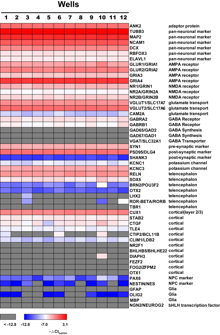
Lowering total tau levels is an attractive therapeutic strategy for Alzheimer's disease and other tauopathies. High-throughput screening in neurons derived from human induced pluripotent stem cells (iPSCs) is a powerful tool to identify tau-targeted therapeutics. However, such screens have been hampered by heterogeneous neuronal production, high cost and low yield, and multi-step differentiation procedures. We engineered an isogenic iPSC line that harbors an inducible neurogenin 2 transgene, a transcription factor that rapidly converts iPSCs to neurons, integrated at the AAVS1 locus. Using a simplified two-step protocol, we differentiated these iPSCs into cortical glutamatergic neurons with minimal well-to-well variability. We developed a robust high-content screening assay to identify tau-lowering compounds in LOPAC and identified adrenergic receptors agonists as a class of compounds that reduce endogenous human tau. These techniques enable the use of human neurons for high-throughput screening of drugs to treat neurodegenerative disease.

Keywords: Alzheimer’s disease; Tau-lowering; adrenergic receptor; frontotemporal dementia; high-content screening; human induced pluripotent stem cells; human neurons; neurodegeneration; neurogenin 2; tau.


Figures

Figure 1
Figure 1
Engineering of i3N iPSCs and Generation of Homogeneous Functional Glutamatergic Neurons by a Simplified Two-Step Procedure (A) Schematic of the targeting of the AAVS1 locus with pUCM.Puro-CAG.rtTA3G-TRE3G.Ngn2 donor vector by TALEN-mediated integration. The third-generation doxycycline-inducible reverse transcriptional activator (rtTA3G) is driven by the CAG promoter and followed by rbGlob polyA tail. Mouse Ngn2 is driven by the tet response element (TRE3G) and followed by SV40 polyA tail. It is oriented tail-to-tail with rtTA3G. Orange boxes are exons of the PPP1R12C gene; gray boxes are regions of homology. PCR1 and PCR2 primers are used for 5′ and 3′ junction PCR screening and generate 1.1-kb and 1.5-kb PCR products, respectively. PCR3 primers (product size, 248 base pairs) are used to detect the nonintegrated allele at the AAVS1 locus. (B) Flow diagram of the two-step procedure for generating i3Neurons. (C) Representative phase-contrast images during the differentiation of i3Neurons. The timeline is the same as shown in (B). Scale bar, 50 μm. (D) Representative images showing immunocytochemical staining for the pan-neuronal marker MAP2, βIII tubulin (TUJ1 antibody), and NeuN in i3Neurons after 4 weeks of differentiation. Nuclei were labeled by Hoechst. Scale bar, 25 μm. (E) Representative images of immunocytochemical staining of mature 8-week-old i3Neurons show tau enrichment (detected with HT7) in an axon identified by the axon initiation segment marker ankyrin G (AnkG). Nuclei were labeled by Hoechst. Scale bar, 25 μm. (F) Representative confocal images of i3Neurons showing immunolabeling of postsynaptic GluR2/3 containing AMPA-type glutamate receptors (red) and the presynaptic vesicular glutamate transporter VGlut1 (green). The colocalization of GluR2/3 and VGlut1 puncta marks glutamatergic synapses formed between i3Neurons. Scale bar, 5 μm. (G) Representative traces of action potentials evoked by 500-ms current step injections at just above the firing threshold (green trace) and at a higher firing frequency (black trace). (H) Spontaneous excitatory postsynaptic currents recorded from an i3N neuron (top) were blocked by CNQX, an AMPA receptor antagonist (bottom). See also Figures S1 and S2.
Figure 2
Figure 2
i3Neurons Show Homogeneous Expression of Glutamatergic Cortical Neuronal Genes Heatmap of RT-qPCR analysis of expression levels of genes listed on the right. Expression levels are normalized to housekeeping gene GAPDH (expressed as −ΔCt) and color coded as shown. mRNA was harvested from 12 random wells of 4-week-old i3Neurons cultured in a 96-well plate. See also Table S1.
Figure 3
Figure 3
Development and Validation of an HCS Assay to Detect Tau Levels in i3Neurons (A) Schematic of the HCS assay optimized to measure cellular tau levels in neurons treated with small-molecule compounds. (B) Representative fluorescence high-content images showing tau (green) and βIII tubulin (white) channels from a background well (left, anti-TUJ1 only) and a control well (right, anti-HT7 and TUJ1). Neurite regions (purple) were traced according to the TUJ1 channel and were applied to the tau channel with the neuronal profiling module of Cellomics software. Scale bar, 10 μm. (C) Representative fluorescence high-content images showing tau (green) and βIII tubulin (white) channels from i3Neurons after 7 days of treatment with control siRNA or human tau siRNA (0.5 or 1 μM). Scale bar, 50 μm. (D) Automated quantification of human tau levels (left) and neurite total length (right) from i3Neurons treated with human tau siRNA. Data are from three independent experiments, total N = 42 per treatment; values are means ± SEM relative to control siRNA. ∗∗∗∗p < 0.0001 compared with control siRNA, STATA mixed model. ####p < 0.0001, STATA mixed model. (E) Automated quantification of human tau levels (left) and neurite total length (right) from i3Neurons treated with 5 mM salicylate, 1 μM YM-01, or 1.5 μM methylene blue for 24–72 hr. Data are from three independent experiments, total N = 42 per treatment; values are means ± SEM relative to DMSO. ∗∗∗p < 0.001, ∗∗∗∗p < 0.0001, compared with DMSO, STATA mixed model; #p < 0.05, ###p < 0.001, ####p < 0.0001, comparison between three time points within each compound treatment, STATA mixed model. See also Figure S3.
Figure 4
Figure 4
Primary Screening and Hit Selection from LOPAC (A) Overview of automated quantification of human tau levels from i3Neurons incubated with compounds from LOPAC at 10 μM for 3 days (green triangles). The library was distributed to three and one-half 384-well plates; each plate contains 16 control wells (DMSO, orange circles) and 16 background wells (blue diamonds). Results are shown as the Z score, calculated based on LOPAC compounds on each plate. Compounds above (yellow triangles) and below (red triangles) the cut-offs of +3 or −3 (dotted lines) are considered hits. (B and C) Hits are ranked by neurite total length (B) and valid nucleus count (C). Results are shown as Z score, calculated based on LOPAC compounds on each plate. Moxonidine and metaproterenol are the top two tau-lowering hits from both rankings.
Figure 5
Figure 5
Activation of α- and β-AR Reduces Total Tau Levels in i3Neurons (A) Representative calibration curve of HT7-Tau5 ultra-sensitive human tau ELISA. Inset shows the assay's limit of quantification (LOQ). (B) 7-day incubation of human tau siRNA significantly lowered total tau levels in i3Neurons. Human tau levels were quantified by HT7-Tau5 ELISA and normalized to protein level. Values are means ± SEM relative to control siRNA. Data are from one experiment, N = 6 wells per treatment. (C–E) Concentration-response curve of moxinidine (C), clonidine (D), and metaproterenol (E) determined by HT7-Tau5 ELISA. Insets show the βIII tubulin level for each concentration as determined by βIII tubulin ELISA. Both tau and βIII tubulin levels are normalized to protein levels. Values are means ± SEM relative to DMSO control. Data are from four independent experiments performed in triplicate (C, N = 12 per concentration) and three independent experiments performed in triplicate (D and E, N = 9 per concentration). p < 0.05, ∗∗p < 0.01, ∗∗∗p < 0.001; STATA mixed model. (F and G) 3-day incubation with moxonidine (30 μM) and the α-AR agonist dexmedetomidine (100 μM) (F) or metaproterenol (30 μM) and the β-AR agonist isoproterenol (30 μM) (G) significantly reduced total tau levels in i3Neurons. The α-AR antagonist atipamezole (100 μM) (F) and the β-AR antagonist propranolol (50 μM) (G) increased total tau levels and abolished the tau-lowering effect of moxonidine (F) or metaproterenol (G), respectively. Human tau levels were quantified by HT7-Tau5 ELISA and normalized to protein level. Values are means ± SEM relative to DMSO control. Data are from three independent experiments performed in triplicate (F, N = 9 per treatment) and four independent experiments performed in triplicate (G, N = 12 per treatment) ∗∗∗p < 0.001, STATA mixed model. See also Figure S4.

References

    1. Abisambra J., Jinwal U.K., Miyata Y., Rogers J., Blair L., Li X., Seguin S.P., Wang L., Jin Y., Bacon J. Allosteric heat shock protein 70 inhibitors rapidly rescue synaptic plasticity deficits by reducing aberrant tau. Biol. Psychiatry. 2013;74:367–374. - PMC - PubMed
    1. Albrecht H., Zbinden P., Rizzi A., Villetti G., Riccardi B., Puccini P., Catinella S., Imbimbo B.P. High throughput screening of beta-amyloid secretion inhibitors using homogenous time-resolved fluorescence. Comb. Chem. High Throughput Screen. 2004;7:745–756. - PubMed
    1. Amamoto R., Arlotta P. Development-inspired reprogramming of the mammalian central nervous system. Science. 2014;343:1239882. - PMC - PubMed
    1. Andorfer C., Acker C.M., Kress Y., Hof P.R., Duff K., Davies P. Cell-cycle reentry and cell death in transgenic mice expressing nonmutant human tau isoforms. J. Neurosci. 2005;25:5446–5454. - PMC - PubMed
    1. Andrews-Zwilling Y., Bien-Ly N., Xu Q., Li G., Bernardo A., Yoon S.Y., Zwilling D., Yan T.X., Chen L., Huang Y. Apolipoprotein E4 causes age- and Tau-dependent impairment of GABAergic interneurons, leading to learning and memory deficits in mice. J. Neurosci. 2010;30:13707–13717. - PMC - PubMed

Publication types

MeSH terms