Review
. 2017 Nov 15;57(11):584-589.
doi: 10.2176/nmc.ra.2017-0030. Epub 2017 Oct 2.

Functional Vascular Anatomy of the Brain

Affiliations
Review

Functional Vascular Anatomy of the Brain

Michihiro Tanaka. Neurol Med Chir (Tokyo). .

Abstract

Functional vascular anatomy is the study of anatomy in its relation to the function that figures out the normal and pathological vascularization of the brain and spinal cord. The mechanism of anatomical variations (e.g. fenestration of the basilar artery, persistent primitive trigeminal artery, and aberrant subclavian artery) can be explained according to the embryological development of the cardiovascular system. The most developmental process is common among the species of the vertebrates from the fish to the mammalian in the early phase of embryo. Thus, it is possible to deduce the reasons of vascular variants in terms of phylogeny. Such an embryological parallelism like the comparative anatomy provides the new insights into the nature of our vascular system. In addition, learning more about the hemodynamic consequence may help to realize the underlying physiopathology of cerebral arterial remodeling and stroke in patients with these vascular variants. This perception may facilitate better understanding of the vascular pathologies and lead to the appropriate decision making not only in the diagnostic work, but also in the interventional procedures. The aim of this study is to introduce the meanings of functional anatomy in the clinical application of vascular diseases and anomalous of the central nervous system.

Keywords: balloon occlusion test; collateral circulation; embryology; functional vascular anatomy; hemodynamic tolerance; phylogeny.


Conflict of interest statement

Conflicts of Interest Disclosure

I declare that I have no conflict of interest.

Figures
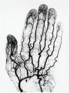
Fig. 1
Fig. 1
Angiography of right radial artery. Note there are numerous collateral circulations. Deep palmar arch is a prominent communicating artery between radial artery and ulnar artery. (arrow). In each segment of the digit, the anastomosis can be observed between two proper palmar digital arteries. These collateral networks are functioning as the security system for the important organ like a distal segment of the finger. The density of the arterial network on the fingertips of thumb is higher than the little finger. This difference corresponds to the density of the Vater-Pacini bodies on the finger.
Fig. 2
Fig. 2
MRA (TOF) showing a lower basilar trunk fenestration. This is the most common variant of the arteries. This fenestration is associated with unfused condition because the basilar artery develops from paired primitive longitudinal neural arteries in terms of functional anatomy.
Fig. 3
Fig. 3
3D rotation angiography showing the paraclinoid aneurysm associated with the intracavernous origin of the ophthalmic artery. It is postulated that there is a relationship between anomalous of ophthalmic artery and formation of paraclinoid aneurysm.
Fig. 4
Fig. 4
Right ICA angiography under left ICA balloon occlusion test in the treatment of traumatic carotid cavernous high flow fistula. Immediate after balloon inflation (A) and three minutes after balloon inflation (B). Note the significant dilatation of anterior communicating artery and left A1 segment. This is a phenomenon of immediate adaptation to left ICA occlusion to compensate the hemodynamic compromise in the area of left MCA cortical territory. This functional caliber change is based on the autoregulation of cerebral arteries.
Fig. 5
Fig. 5
(A) Left internal carotid angiography shows fusiform aneurysm locating distal PCA (P3–P4 segment). (B) Through the fetal type of posterior communicating artery, a microballoon catheter was navigated and inflated at the P3 segment that is just proximal to the aneurysm. (C) Left internal carotid angiography under balloon occlusion revealed a retrograde filling of P4 segment through the parieto-occipital branch of PCA. This functional test indicated sufficient collateral circulation through the leptomeningeal anastomosis from MCA cortical territory. It enables the internal trapping of the aneurysm including parent artery without hemodynamic compromise in the territory of PCA.
Fig. 6
Fig. 6
(A, B) 3D-RA frontal view corresponding to the DSA showing retrograde filling to the parieto-occipital artery (arrow). The use of automated vessel analysis software allows determination of the entire course of the vessel in the region of interest. Note the blue color line on 3D-RA delineating the major collateral pathway of leptomeningeal anastomosis through the angular artery of MCA.
Fig. 7
Fig. 7
3D-RA lateral view (A) and the schematic representation by Zülch (B) demonstrating the potential anastomosis at the level of the cortical artery.

References

    1. Lasjaunias P, Berenstein A, ter Brugge KG: Clinical Vascular Anatomy and Variations. Berlin, Heidelberg, Springer Berlin Heidelberg, 2001.
    1. Sloffer CA, Lanzino G: Historical vignette. Dominique Anel: father of the hunterian ligation? J Neurosurg 104: 626– 629, 2006. - PubMed
    1. Padget DH: The development of the cranial arteries in the human embryo. Contr Embryol 32: 205– 261, 1948.
    1. Tanaka M, Kikuchi Y, Ouchi T: Neuroradiological analysis of 23 cases of basilar artery fenestration based on 2280 cases of MR angiographies. Interv Neuroradiol 12 (Suppl 1): 39–44, 2006. - PMC - PubMed
    1. Black SP, Ansbacher LE: Saccular aneurysm associated with segmental duplication of the basilar artery. A morphological study. J Neurosurg 61: 1005– 1008, 1984. - PubMed