. 2017 Sep-Oct;51(5):493-504.
doi: 10.4103/ortho.IJOrtho_164_17.

Current Concepts in the Management of Patellar Instability

Affiliations

Current Concepts in the Management of Patellar Instability

Michael S Laidlaw et al. Indian J Orthop. 2017 Sep-Oct.

Abstract

Patellar instability is a common presenting clinical entity in the field of orthopedics. This not only can occur from baseline morphologic variability within the patellofemoral articulation and alignment, but also from traumatic injury. While conservative management is many times employed early in the treatment course, symptomatic patellar instability can persist. This article reviews the available indexed published literature regarding patellar instability. Given the debilitating nature of this condition and the long term sequelae that can evolve from its lack of adequate recognition and treatment, this article details the most current methods in the evaluation of this entity as well as reviews the most up-to-date surgical treatment regimens that are available to address this condition.

Keywords: Medial patellofemoral ligament; Patella; osteotomy; patella alta; patella femoral pain syndrome; patellar dislocation; patellar instability; tibial tubercle osteotomy; tibial tubercle-trochlear groove; trochlear dysplasia; trochleoplasty.


Conflict of interest statement

Dr. Diduch is a consultant for Depuy Synthes and receives royalties from Smith and Nephew; however, for the purposes of this review article, there are no gross conflicts of interest.

Figures
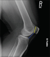
Figure 1
Figure 1
A lateral radiograph of right knee joint showing the Caton–Deschamps index measured as the ratio of the blue line/yellow line (blue line distance measured from the tibial plateau to the inferior articular margin of the patella. Yellow line distance measured from the superior to inferior extent of patellar articular margin)
Figure 2
Figure 2
(a) Standard axial patellar radiograph view of knee in 30° of flexion. (b) Computed tomography arthrogram axial image of the same knee in full extension revealing the presence of a supratrochlear spur
Figure 3
Figure 3
Trochlear dysplasia classification according to D. Dejour. In Type A, the crossing sign is present. Type B features include the crossing sign and trochlear spur. In Type C there is a crossing sign and the double-contour sign. Type D combines the crossing sign, supratrochlear spur and double-contour sign (with permission of Springer: Dejour D, Saggin P. The sulcus deepening trochleoplasty-the Lyon's procedure. Int Orthop 2010;34:313 Figure 2)
Figure 4
Figure 4
Axial computed tomography scan image left knee showing the tibial tubercle-trochlear groove has been measured at 22.11. This is measured by a perpendicular line drawn from the posterior femoral condylar axis through the bisecting point of the trochlear groove. This line is then superimposed onto the axial image of the midpoint of the tibial tubercle. The line drawn perpendicular to this trochlear groove line to the bisecting point of the tibial tubercle represents the tibial tubercle-trochlear groove distance
Figure 5
Figure 5
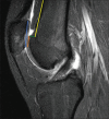
Sagittal magnetic resonance imaging T2W image measures the patellar trochlear index at 40%. The blue line corresponds to the superior to inferior most patellar articular cartilage. A right angle to that inferior most patellar cartilage is used to then measure a parallel line marking the length of corresponding trochlear cartilage to its superior-most extent. Patellar trochlear index = Orange/Blue ×100%. Furthermore, the yellow line marks the anterior femoral cortex and the supratrochlear spur is measured by the green line, which in this case is 6 mm in height
Figure 6
Figure 6
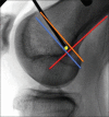
A perfect intraoperative C-arm lateral image of a left knee with complete posterior femoral condyle overlap. Schöttle's point is detailed as a yellow dot, noted as 1 mm anterior to the posterior cortex extension line (red line), 2.5 mm distal to the posterior origin of the medial femoral condyle (orange line), and proximal to the level of the posterior point of Blumensaat's line (blue line)
Figure 7
Figure 7
Intraoperative photos of left knee. (a) Pretrochleoplasty: Dejour B trochlea with nonexistent sulcus angle and supratrochlear spur. (b) Posttrochleoplasty: Note the medial/lateral shingle creation, with the reduction maintained by suture anchors loaded with #2 absorbable suture, and the restored sulcus with slight lateralization of the trochlear groove
Figure 8
Figure 8
Intraoperative lateral C-arm images of a left knee: (a) Note the distalized feathered shingle (blue arrow) used to address mild patella alta. (b) The step-cut is performed to address severe patella alta (short red arrow reveals the distalization of the osteotomy shingle and the long red arrow shows the bone segment that was cut distally and transferred proximally as autogenous bone grafting)

Similar articles

Cited by

References

    1. Fithian DC, Paxton EW, Stone ML, Silva P, Davis DK, Elias DA, et al. Epidemiology and natural history of acute patellar dislocation. Am J Sports Med. 2004;32:1114–21. - PubMed
    1. Arendt EA, Fithian DC, Cohen E. Current concepts of lateral patella dislocation. Clin Sports Med. 2002;21:499–519. - PubMed
    1. Atkin DM, Fithian DC, Marangi KS, Stone ML, Dobson BE, Mendelsohn C, et al. Characteristics of patients with primary acute lateral patellar dislocation and their recovery within the first 6 months of injury. Am J Sports Med. 2000;28:472–9. - PubMed
    1. Nietosvaara Y, Aalto K, Kallio PE. Acute patellar dislocation in children: Incidence and associated osteochondral fractures. J Pediatr Orthop. 1994;14:513–5. - PubMed
    1. Elias DA, White LM, Fithian DC. Acute lateral patellar dislocation at MR imaging: Injury patterns of medial patellar soft-tissue restraints and osteochondral injuries of the inferomedial patella. Radiology. 2002;225:736–43. - PubMed

LinkOut - more resources