

Primary Cutaneous Endometriosis of Umbilicus
- PMID: 28966521
- PMCID: PMC5597658
- DOI: 10.5021/ad.2017.29.5.621
Primary Cutaneous Endometriosis of Umbilicus
Abstract
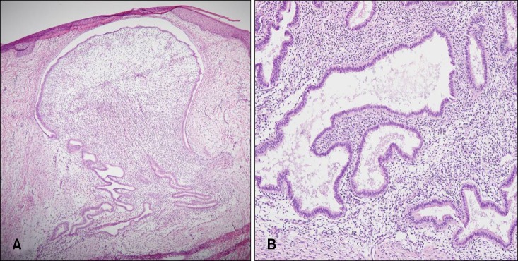
Cutaneous endometriosis is defined by the presence of endometrial glands and/or stroma in skin and represents less than 1% of all ectopic endometrium. Cutaneous endometriosis is classified as primary and secondary. Primary cutaneous endometriosis appears without a prior surgical history and secondary cutaneous endometriosis mostly occurs at surgical scar tissue after abdominal operations. The most widely accepted pathogenesis of secondary endometriosis is the iatrogenic implantation of endometrial cells after surgery, such as laparoscopic procedures. However, the pathogenesis of primary endometriosis is still unknown. Umbilical endometriosis is composed only 0.4% to 4.0% of all endometriosis, however, umbilicus is the most common site of primary cutaneous endometriosis. A 38-year-old women presented with solitary 2.5×2.0-cm-sized purple to brown colored painful nodule on the umbilicus since 2 years ago. The patient had no history of surgical procedures. The skin lesion became swollen with spontaneous bleeding during menstruation. The skin lesion was diagnosed as a keloid at private hospital and has been treated with lesional injection of steroid for several times but there was no improvement. Imaging studies showed an enhancing umbilical mass without connection to internal organs. Biopsy specimen showed the several dilated glandular structures in dermis. They were surrounded by endometrial-type stroma and perivascular infiltration of lymphocytes. The patient was diagnosed as primary cutaneous endometriosis and skin lesion was removed by complete wide excision without recurrence. We report an interesting and rare case of primary umbilical endometriosis mistaken for a keloid and review the literatures.
Keywords: Cutaneous endometriosis; Endometriosis of umbilicus; Primary cutaneous endometriosis; Umbilical endometriosis.
Conflict of interest statement
CONFLICTS OF INTEREST: The authors have nothing to disclose.
Figures

References
-
- Minaidou E, Polymeris A, Vassiliou J, Kondi-Paphiti A, Karoutsou E, Katafygiotis P, et al. Primary umbilical endometriosis: case report and literature review. Clin Exp Obstet Gynecol. 2012;39:562–564. - PubMed
-
- Yuen JS, Chow PK, Koong HN, Ho JM, Girija R. Unusual sites (thorax and umbilical hernial sac) of endometriosis. J R Coll Surg Edinb. 2001;46:313–315. - PubMed
-
- Fernández-Aceñero MJ, Córdova S. Cutaneous endometriosis: review of 15 cases diagnosed at a single institution. Arch Gynecol Obstet. 2011;283:1041–1044. - PubMed
-
- Weng CS, Yang YC. Images in clinical medicine. Villar's nodule--umbilical endometriosis. N Engl J Med. 2011;364:e45. - PubMed
Publication types
LinkOut - more resources
Full Text Sources
Other Literature Sources
