

Diagnostic application of clinical exome sequencing in Leber congenital amaurosis
- PMID: 28966547
- PMCID: PMC5610811
Diagnostic application of clinical exome sequencing in Leber congenital amaurosis
Abstract
Purpose: Leber congenital amaurosis (LCA) is a hereditary retinal dystrophy with wide genetic heterogeneity. Next-generation sequencing (NGS) targeting multiple genes can be a good option for the diagnosis of LCA, and we tested a clinical exome panel in patients with LCA.
Methods: A total of nine unrelated Korean patients with LCA were sequenced using the Illumina TruSight One panel, which targets 4,813 clinically associated genes, followed by confirmation using Sanger sequencing. Patients' clinical information and familial study results were obtained and used for comprehensive interpretation.
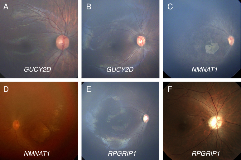
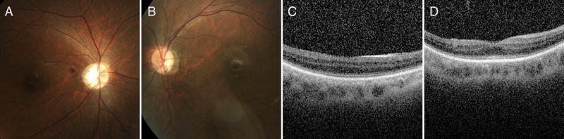
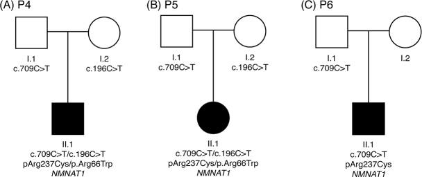
Results: In all nine patients, we identified pathogenic variations in LCA-associated genes: NMNAT1 (n=3), GUCY2D (n=2), RPGRIP1 (n=2), CRX (n=1), and CEP290 or SPATA7. Six patients had one or two mutations in accordance with inheritance patterns, all consistent with clinical phenotypes. Two patients had only one pathogenic mutation in recessive genes (NMNAT1 and RPGRIP1), and the clinical features were specific to disorders associated with those genes. Six patients were solved for genetic causes, and it remains unclear for three patients with the clinical exome panel. With subsequent targeted panel sequencing with 113 genes associated with infantile nystagmus syndrome, a likely pathogenic allele in CEP290 was detected in one patient. Interestingly, one pathogenic variant (p.Arg237Cys) in NMNAT1 was present in three patients, and it had a high allele frequency (0.24%) in the general Korean population, suggesting that NMNAT1 could be a major gene responsible for LCA in Koreans.
Conclusions: We confirmed that a commercial clinical exome panel can be effectively used in the diagnosis of LCA. Careful interpretation and clinical correlation could promote the successful implementation of clinical exome panels in routine diagnoses of retinal dystrophies, including LCA.
Figures
Similar articles
-
Copy number variations and multiallelic variants in Korean patients with Leber congenital amaurosis.Mol Vis. 2020 Feb 24;26:26-35. eCollection 2020. Mol Vis. 2020. PMID: 32165824 Free PMC article.
-
The genetic profile of Leber congenital amaurosis in an Australian cohort.Mol Genet Genomic Med. 2017 Nov;5(6):652-667. doi: 10.1002/mgg3.321. Epub 2017 Aug 22. Mol Genet Genomic Med. 2017. PMID: 29178642 Free PMC article.
-
Molecular background of Leber congenital amaurosis in a Polish cohort of patients-novel variants discovered by NGS.J Appl Genet. 2023 Feb;64(1):89-104. doi: 10.1007/s13353-022-00733-9. Epub 2022 Nov 12. J Appl Genet. 2023. PMID: 36369640 Free PMC article.
-
Clinical and genetic findings in a family with NMNAT1-associated Leber congenital amaurosis: case report and review of the literature.Graefes Arch Clin Exp Ophthalmol. 2015 Dec;253(12):2239-46. doi: 10.1007/s00417-015-3174-0. Epub 2015 Oct 13. Graefes Arch Clin Exp Ophthalmol. 2015. PMID: 26464178 Review.
-
Clinical course of a Japanese girl with Leber congenital amaurosis associated with a novel nonsense pathogenic variant in NMNAT1: a case report and mini review.Ophthalmic Genet. 2022 Jun;43(3):400-408. doi: 10.1080/13816810.2021.2023195. Epub 2022 Jan 13. Ophthalmic Genet. 2022. PMID: 35026968 Review.
Cited by
-
Molecular Diagnosis of 34 Japanese Families with Leber Congenital Amaurosis Using Targeted Next Generation Sequencing.Sci Rep. 2018 May 29;8(1):8279. doi: 10.1038/s41598-018-26524-z. Sci Rep. 2018. PMID: 29844330 Free PMC article.
-
Copy number variations and multiallelic variants in Korean patients with Leber congenital amaurosis.Mol Vis. 2020 Feb 24;26:26-35. eCollection 2020. Mol Vis. 2020. PMID: 32165824 Free PMC article.
-
Clinical features and genetic spectrum of NMNAT1-associated retinal degeneration.Eye (Lond). 2022 Dec;36(12):2279-2285. doi: 10.1038/s41433-021-01853-y. Epub 2021 Nov 26. Eye (Lond). 2022. PMID: 34837036 Free PMC article.
-
Accuracy of Next-Generation Sequencing for Molecular Diagnosis in Patients With Infantile Nystagmus Syndrome.JAMA Ophthalmol. 2017 Dec 1;135(12):1376-1385. doi: 10.1001/jamaophthalmol.2017.4859. JAMA Ophthalmol. 2017. PMID: 29145603 Free PMC article.
-
Voretigene Neparvovec for the Treatment of RPE65-associated Retinal Dystrophy: Consensus and Recommendations from the Korea RPE65-IRD Consensus Paper Committee.Korean J Ophthalmol. 2023 Apr;37(2):166-186. doi: 10.3341/kjo.2023.0008. Epub 2023 Mar 23. Korean J Ophthalmol. 2023. PMID: 36950921 Free PMC article.
References
-
- De Laey JJ. Leber’s congenital amaurosis. Bull Soc Belge Ophtalmol. 1991;241:41–50. - PubMed
-
- den Hollander AI, Roepman R, Koenekoop RK, Cremers FP. Leber congenital amaurosis: genes, proteins and disease mechanisms. Prog Retin Eye Res. 2008;27:391–419. - PubMed
-
- Schappert-Kimmijser J, Henkes HE, Van Den Bosch J. Amaurosis congenita (Leber). AMA Arch Opthalmol. 1959;61:211–8. - PubMed
-
- Estrada-Cuzcano A, Koenekoop RK, Coppieters F, Kohl S, Lopez I, Collin RW, De Baere EB, Roeleveld D, Marek J, Bernd A, Rohrschneider K, van den Born LI, Meire F, Maumenee IH, Jacobson SG, Hoyng CB, Zrenner E, Cremers FP, den Hollander AI. IQCB1 mutations in patients with leber congenital amaurosis. Invest Ophthalmol Vis Sci. 2011;52:834–9. - PubMed
-
- Koenekoop RK, Wang H, Majewski J, Wang X, Lopez I, Ren H, Chen Y, Li Y, Fishman GA, Genead M, Schwartzentruber J, Solanki N, Traboulsi EI, Cheng J, Logan CV, McKibbin M, Hayward BE, Parry DA, Johnson CA, Nageeb M, Poulter JA, Mohamed MD, Jafri H, Rashid Y, Taylor GR, Keser V, Mardon G, Xu H, Inglehearn CF, Fu Q, Toomes C, Chen R. Mutations in NMNAT1 cause Leber congenital amaurosis and identify a new disease pathway for retinal degeneration. Nat Genet. 2012;44:1035–9. - PMC - PubMed
MeSH terms
Substances
LinkOut - more resources
Full Text Sources
Research Materials
