

A novel triple immunoenzyme staining enables simultaneous identification of all muscle fiber types on a single skeletal muscle cryosection from normal, denervated or reinnervated rats
- PMID: 28966653
- PMCID: PMC5607833
- DOI: 10.4103/1673-5374.213560
A novel triple immunoenzyme staining enables simultaneous identification of all muscle fiber types on a single skeletal muscle cryosection from normal, denervated or reinnervated rats
Abstract
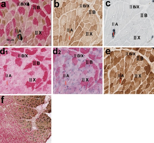
Triple immunofluorescence staining has recently been developed to simultaneously identify all muscle fibers on a single cryosection which is helpful for clinical and basic research, but it has disadvantages such as fast photobleaching and unclear outlines of muscle fibers. Triple immunoenzyme staining (TIE) is likely to avoid these disadvantages. In this study, we aimed to establish a sensitive and specific TIE technique to identify fiber types in normal, denervated, and reinnervated rat muscles, and to develop a systematic sampling method for muscle fiber quantification. Tibialis anterior and soleus from normal, denervated, and reinnervated Lewis rat hind limbs were used. Five consecutive cryosections were cut from each muscle, including one for TIE and four for single immunoenzyme staining (SIE). The TIE was performed using the polymerized reporter enzyme staining system for the first two antigens (A4.74 for MyHC-IIA, BA-F8 for MyHC-I) and alkaline phosphatase staining system for the third antigen (BF-F3 for MyHC-IIB), followed by corresponding detective systems and respective chromogens. The type of muscle fibers was quantified by systematic sampling at 12.5%, 25%, 33% and 50% of all muscle fibers, and was compared with that acquired from counting all the fibers (100%). All muscle fiber phenotypes, including pure and hybrid, could be simultaneously identified on a single TIE cryosection with clear outlines. The fiber types on TIE slides matched well with their respective counterpart on the consecutive SIE slides with a 95% match rate. Systematic sampling of 12.5% fibers could represent the true fiber type distribution of the entire muscle section. Our results suggest that novel TIE can effectively visualize fiber types in normal, denervated or reinnervated rat muscles.
Keywords: immunohistochemistry; muscle fiber phenotyping; myosin heavy chain; nerve regeneration; neural regeneration; rats; triple immunoenzyme staining.
Conflict of interest statement
Conflicts of interest: None declared.
Figures


References
-
- Armstrong RB, Phelps RO. Muscle fiber type composition of the rat hindlimb. Am J Anat. 1984;171:259–272. - PubMed
-
- Bigard AX, Serrurier B, Merino D, Lienhard F, Berthelot M, Guezennec CY. Myosin heavy chain composition of regenerated soleus muscles during hindlimb suspension. Acta Physiol Scand. 1997;161:23–30. - PubMed
-
- Bobinac D, Malnar-Dragojevic D, Bajek S, Soic-Vranic T, Jerkovic R. Muscle fiber type composition and morphometric properties of denervated rat extensor digitorum longus muscle. Croat Med J. 2000;41:294–297. - PubMed
-
- Claassen E, Boorsma DM, Kors N, Van Rooijen N. Double-enzyme conjugates, producing an intermediate color, for simultaneous and direct detection of three different intracellular immunoglobulin determinants with only two enzymes. J Histochem Cytochem. 1986;34:423–428. - PubMed
Grants and funding
LinkOut - more resources
Full Text Sources
Other Literature Sources
Miscellaneous
