. 2017 Oct 1;8(5):519-530.
doi: 10.14336/AD.2017.0710. eCollection 2017 Oct.

Aging Systemic Milieu Impairs Outcome after Ischemic Stroke in Rats

Affiliations

Aging Systemic Milieu Impairs Outcome after Ischemic Stroke in Rats

Mengxiong Pan et al. Aging Dis. .

Abstract

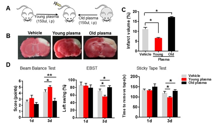
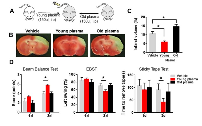
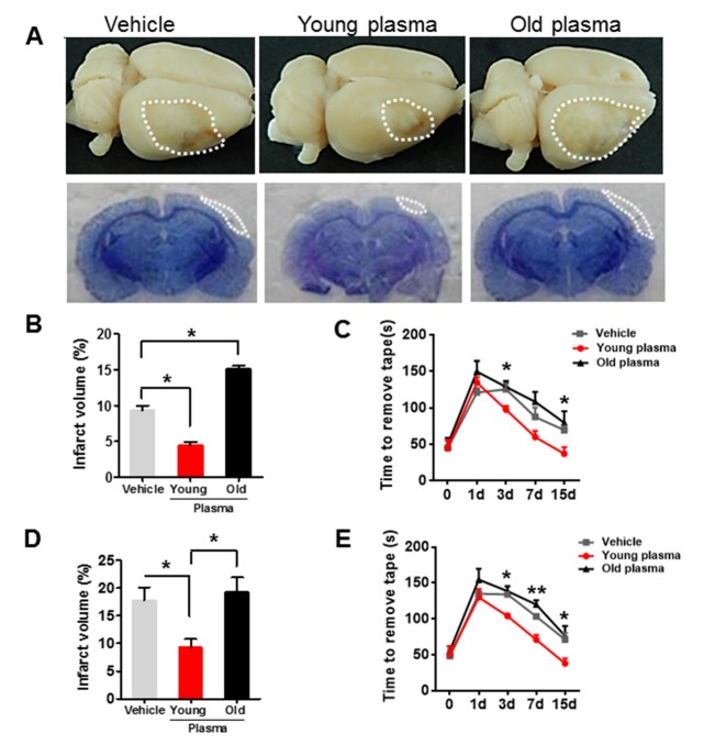
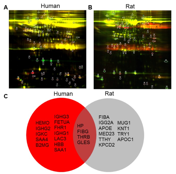
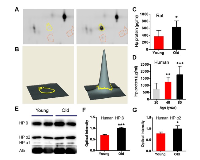
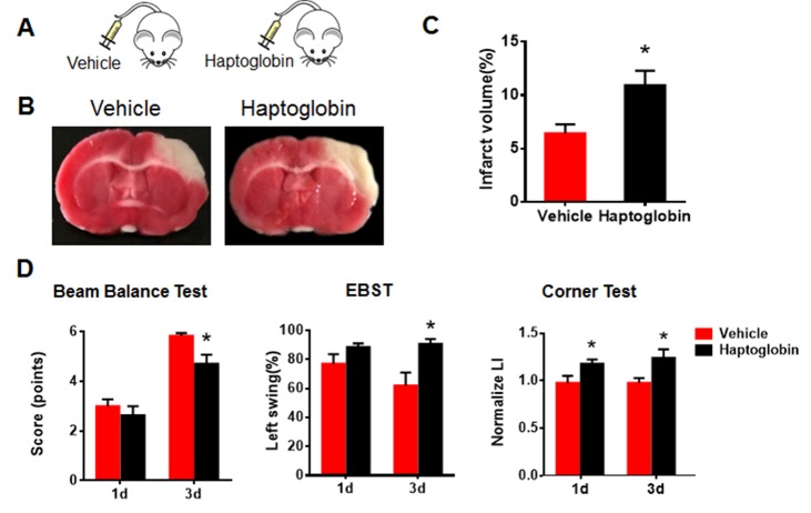
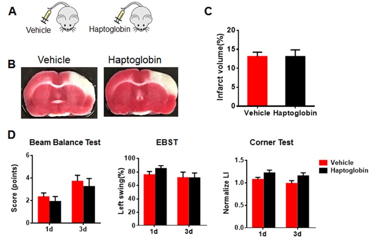
Compelling evidence indicates that factors in the blood can profoundly reverse aging-related impairments, as exposure of aged mice to young blood rejuvenates adult stem cell function, improves cognition, and ameliorates cardiac hypertrophy. Systemic factors from mice can also extend the life span of a partner exposed to a lethal treatment or disease. These findings suggest that the systemic milieu of a healthy young partner may be beneficial for an aged organism. However, it is unknown whether a healthy young systemic milieu can improve functional recovery after ischemic stroke. Intraperitoneal administration of young plasma into aged rats after ischemic stroke induced by distal middle cerebral artery occlusion (dMCAO) reduced infarct volume and motor impairment, compared with vehicle group. On the contrary, intraperitoneal administration of plasma from aged rats into young ischemic rats worsened brain injury and motor deficits. Using a proteomic approach, we found that haptoglobin levels were significantly increased in serum of aged rats and that intraperitoneal administration of haptoglobin impaired outcome after ischemic stroke in young rats. Our data suggest that the aging systemic milieu plays a critical role in functional outcome after ischemic stroke.

Keywords: haptoglobin; ischemic stroke; outcome; plasma; systemic milieu.


Figures

Figure 1.
Figure 1.. Administration of old plasma into young ischemic rats worsensyoungyoungwrosew were outcomeafter ischemic stroke
(A) Schematic illustrating intraperitonealinjection of young (2-month-old) or old (22-month-old) plasma into young ischemic rats 1 hr after onset of ischemia, twice per day for 3 days. (B) Infarct areas (white) in TTC-(red) stained coronal brain section from vehicle, young and old plasma-treated rats at 3 days after stroke. (C) Quantitative analysis of infarct volume in vehicle, young and old plasma-treated rats at 3 days after ischemic stroke. (D) Motor function deficits of young ischemic rats were assessed by beam balance test, EBST and sticky tape test on the 1st and 3rd day after administration with vehicle, young, or old rat plasma. Values presented as mean ± SEM. *P<0.05; **P<0.01, compared with vehicle-treated group. N=7-12 per group.
Figure 2.
Figure 2.. Young plasma reduces youngyounginfarct volume and improves neurobehavioral deficits in aged ischemic rats after stroke
(A) Schematic illustrating intraperitonealinjection of young (2-month-old) or old (22-month-old) plasma into aged ischemic rats 1 hr after onset of ischemia. (B) Representative images of TTC staining in cerebral sections of vehicle, young and old plasma-treated group, twice per day for 3 days after stroke. (C) Infarct volume (expressed as percentage whole brain volume) in vehicle, young and old plasma-treated aged rats 3 days after stroke. (D) Neurobehavioral tests includingbeam balance test, EBST and sticky tape test were performedon the 1st and 3rd day after injection with vehicle, young, or old rat plasma into aged rats. Values presented as mean ± SEM. *P<0.05, compared with vehicle-treated group. N=7-12 per group.
Figure 3.
Figure 3.. Old plasma administration inhibits long-term recovery after experimental stroke in young ischemic rats
(A) Representative images of lesion volume at low magnification view (top panel) and H&E-stained coronal brain sections (bottom panel) in vehicle, young plasma and old plasma-treated young rats 15 days after stroke. (B) Quantitative analysis of infarct volume in vehicle, young and old plasma-treated young rats 15 days after ischemic stroke. (C) Sticky tape test scores in young rats at 1, 3, 7 and 15 days after treatment with vehicle, young plasma and old plasma. (D) Quantitative analysis of infarct volume in vehicle, young and old plasma-treated aged rats 15 days after ischemic stroke. (E) Sticky tape test scores in aged rats at 1, 3, 7 and 15 days after treatment with vehicle, young plasma and old plasma. Values presented as mean ± SEM. *P < 0.05; **P < 0.01, compared with vehicle-treated group. N=7-12 per group.
Figure 4.
Figure 4.. Differentially expressed proteins identified from the 2D-DIGE profiling of young and old plasma of human and rat
(A) Protein from rat young plasma was labeled with Cy3 (green), protein from rat old plasma was labeled with Cy5 (red), and samples were mixed prior to 2-dimensional PAGE (horizontal axis, pI; vertical axis, Mr). (B) Cy3 (green) was used to label young human plasma and Cy5 was used to label old human plasma. N=10/group. (C) Venn diagram of differentially expressed protein spots identified from the 2D-DIGE profiling of young and old human (red) and rat plasma (grey). In the brown intersection are shown four factors changed in both proteomic screens.
Figure 5.
Figure 5.. Circulation level of haptoglobin is significantly increased in both old rat and human plasma
(A) DeCyder Analysis shows the location of a 2D-DIGE gel with yellow line pointing to protein spots, identified as haptoglobin, that was differentially expressed across age, >72.5 change in abundance. (B) 3-D view simulation of a close-up of the region of 2D-DIGE gel image, and the associated graph of representative up-regulated haptoglobin during aging. (C) ELISA shows that haptoglobin was increased in young and old rat plasma. N=7 rats per group. (D) ELISA shows that haptoglobin concentration in the plasma of 20-, 40-, and 80-years-old human subjects. (E) Western blot analysis confirms that increased level of haptoglobin α1, α2 and β in old human plasma. (F and G) Relative optical intensity of haptoglobin β and α2. Data were from 10 per group. Values were presented as mean ± SEM. *P < 0.05, **P < 0.01, ***P < 0.001.
Figure 6.
Figure 6.. Systemic exposure to haptoglobin increases infarct volume and deteriorates neurobehavioral deficits in young ischemic rats
(A) Schematic of young rats injected intraperitoneally with haptoglobin (0.5 g/L in 150 µl) or vehicle 1 hr after ischemic stroke. (B) Representative images of TTC staining in coronal brain sections of vehicle- or haptoglobin-treated young ischemic rats. (C) Quantitative analysis of infarct volume in vehicle- and haptoglobin-treated young rats 3 days after ischemic stroke. (C) Neurobehavioral deficitswere determined by beam balance test, EBST and corner test in young ischemic rats after injection with vehicle or haptoglobin. Values presented as mean ± SEM. *P< 0.05, **P< 0.01. N=7-12 per group.
Figure 7.
Figure 7.. Systemic exposure to haptoglobin affect infarct volume and neurobehavioral deficits in aged ischemic rats
(A) Schematic of aged rats injected intraperitoneally with haptoglobin (0.5 g/L in 150 µl) or vehicle 1 hr after ischemic stroke. (B) Representative images of TTC staining in coronal brain sections of vehicle- or haptoglobin-treated aged ischemic rats. (C) Quantitative analysis of infarct volume in vehicle- and haptoglobin-treated aged rats 3 days after ischemic stroke. (C) Neurobehavioral deficits were determined by beam balance test, EBST and corner test in aged ischemic rats after injection with vehicle or haptoglobin. Values presented as mean ± SEM. N=8-14 per group.

References

    1. Ramirez-Lassepas M (1998). Stroke and the aging of the brain and the arteries. Geriatrics, 53: S44-48. - PubMed
    1. Villeda SA, Plambeck KE, Middeldorp J, Castellano JM, Mosher KI, Luo J, et al. (2014). Young blood reverses age-related impairments in cognitive function and synaptic plasticity in mice. Nat Med, 20: 659-663 - PMC - PubMed
    1. Loffredo FS, Steinhauser ML, Jay SM, Gannon J, Pancoast JR, Yalamanchi P, et al. (2013). Growth differentiation factor 11 is a circulating factor that reverses age-related cardiac hypertrophy. Cell, 153: 828-839 - PMC - PubMed
    1. Conboy IM, Conboy MJ, Wagers AJ, Girma ER, Weissman IL, Rando TA (2005). Rejuvenation of aged progenitor cells by exposure to a young systemic environment. Nature, 433: 760-764 - PubMed
    1. Brack AS, Conboy MJ, Roy S, Lee M, Kuo CJ, Keller C, et al. (2007). Increased Wnt signaling during aging alters muscle stem cell fate and increases fibrosis. Science, 317: 807-810 - PubMed

LinkOut - more resources