. 2017;4(2):e1566.
doi: 10.14800/ccm.1566. Epub 2017 Jul 10.

Brain derived neutrophic factor (BDNF) coordinates lympho-vascular metastasis through a fibroblast-governed paracrine axis in the tumor microenvironment

Affiliations

Brain derived neutrophic factor (BDNF) coordinates lympho-vascular metastasis through a fibroblast-governed paracrine axis in the tumor microenvironment

Tilahun Jiffar et al. Cancer Cell Microenviron. 2017.

Abstract

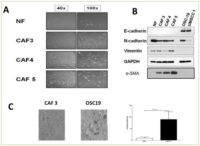
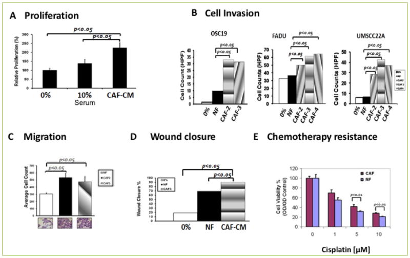
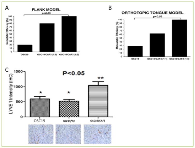
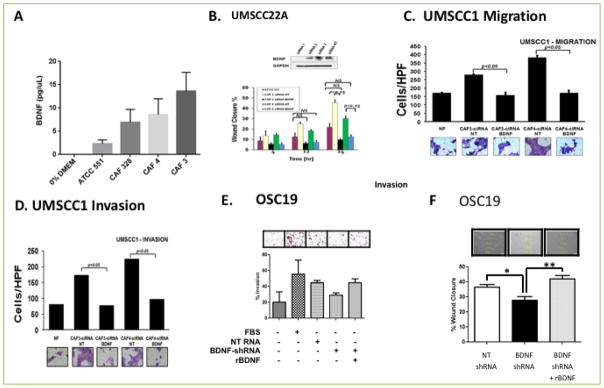
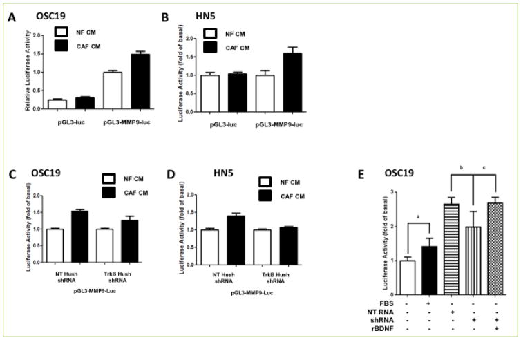
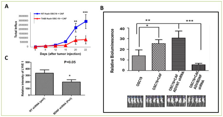
It has long been known that the tumor microenvironment contributes to the proliferation and survival of neoplasms through the constant interaction with the stromal and immune compartments. In this investigation, we explored the role of cancer-associated fibroblasts (CAFs) in the regulation of the tumor microenvironment in head and neck squamous cell carcinoma (HNSCC) though a complex intercellular BDNF-TrkB signaling system. Our studies show that conditioned media derived from patient-derived CAFs promoted HNSCC cell proliferation, in vitro cell migration, cell invasion and chemotherapy resistance, compared to normal fibroblasts. Furthermore, examination of the in vivo impact of CAF pathophysiology in the tumor microenvironment in animal xenograft models revealed that HNSCC cell lines in combination with CAFs promoted tumor growth and increased incidence of lymphovascular metastasis as compared to injection of tumor cells or CAF cells alone. Using pharmacological and genetic alterations, we mechanistically demonstrate the critical importance of BDNF-TrkB signaling in the tumor microenvironment. These investigations further support the rationale for BDNF/TRKB targeted therapy against in the treatment of HNSCC.


Conflict of interest statement

Conflicting interests The authors have declared that no conflict of interests exist.

Figures

Figure 1
Figure 1. Cancer Associated Fibroblasts (CAFs) show morphological and biochemical characteristics of mesenchymal cells
A. Normal fibroblasts and CAFs were allowed to grow in 10% DMEM media. CAFs show distinct morphology from normal fibroblasts. B. Western blot analysis of CAFs and epithelial cells showing mesenchymal markers in CAFs. C. Soft agar assay was used to detect the anchorage independent growth of CAFs and HNSCC cell lines. CAFs and HNSCC cells were suspended in 0.3% of top agar and added to a six-well plate containing a 0.6% solidified agar and incubated for 10 days to investigate anchorage independent colony formation. The gels were then stained with crystal violet stains and colonies lager than 100um were counted under a steriomicroscope. Data represent means ± S.E.M. Significance was determined by Student t-test.
Figure 2
Figure 2. CAF potentiates HNSCC progression including Cell proliferation, migration, invasion, and chemotherapy resistance
A. MTT assay showing a significant (p<0.05) induction of proliferation by CAF conditioned medium in HNSCC cell lines (P<0.05). B. Transwell co-culture of HNSCC cell lines with CAFs showed a significant increase in cell invasion of three HNSCC cell lines, OSC19, FADU, and UMSCC22A when compared to normal fibroblasts (P<0.05). C. HNSCC Cells line were co-cultured with/without CAF and migrating cells were counted. CAF induced significant migration (P<0.05). D. HNSCC cell line was grown to confluency in six well plates and wound scratch was made across the center of the wells. Cells were allowed to heal in the presence of conditioned medium from either of normal fibroblast (NF) or CAF. The distance between the two edges was measured at 0 time and 24 Hrs. Conditioned media from CAFs induced wound closure at a significantly faster rate (P<0.05) than cells incubated with CM from NF. E. HNSCC cell lines were incubated in the presence or absence of CM from NF or CAF in an increasing concentration of cisplatin. HNSCC cells incubated with CM from CAFs were more resistant than those incubated with CM from NF at higher concentration of cisplatin (P<0.05).
Figure 3
Figure 3. CAFs increase metastatic efficiency of HNSCC in mouse xenograft models
A–B. Mice were injected with a suspension of OSC19, OSC19 and CAF (1:1) or OSC19 and CAF (1:5) in matrigel on the flank (A) or on the tongue (B) and maintained until the tumor growth reached 15mm as recommended by the institutional policy. Mice were sacrificed and adjoining lymph nodes and lungs were excised and processed for histological examination. Results indicated CAFs promoted metastatic efficiency (p<0.05) both in the flank model and the tongue model. (C) Immunohistochemical staining for LYVE1, a lymphangiogenic marker, was performed to measure the role of BDNF in potentiation of lymphangiogenesis in mice injected with OSC19, OSC19 and CAF transfected with vector or OSC19 and CAF transfected with BDNF construct. Overexpression of BDNF in CAFs significantly increased lymphangiogenic network as measured by LYVE1 intensity, p<0.05.
Figure 4
Figure 4. CAFs promote tumor cell migration, invasion and progression by activating the TRK-B-BDNF signaling axis
A. ELISA was performed on supernatants derived from normal fibroblasts or CAFS for expression of BDNF and showed that there was a significantly higher level of BDNF secretion in CAFs (p<0.05). B HNCC cell line was grown to confluency and scratched for generation of wound. Wound healing was measured at 4, 12 and 24 Hrs in the presence of either NF or CAFs treated with non-targeting (NT) siRNA or BDNF siRNA. Wound healing was significantly inhibited in the presence of BDNF siRNA (P<0.05). C–D. Genetic suppression of BDNF in CAFs by using BDNF siRNA significantly reduces cellular migration (C) and Cellular invasion (D) in matrigel invasion assay (p<0.05). E. Matrigel invasion assay was performed on HNSCC lines incubated in medium derived from CAFs transfected with vector containing NT shRNA, BDNF shRNA or BDNF shRNA and recombinant BDNF. Addition of exogenous BDNF restored invasion of HNSCC cell line. F. HNSCC cells were grown to confluency in six well plates. Wound was generated by pipet tips along the diameter of the well. Reconstitution of exogenous BDNF (100nM) in the media restored wound healing in HNSCC cell line incubated with CM derived from CAFs transfected with NT shRNA or BDNF shRNA, p<0.05.
Figure 5
Figure 5. BDNF/TrkB system mediates CAF-induced MMP9 expression in HNSCC cell lines
A–B. HNSCC cell lines were transfected with either empty vector (PGL3-Luc) or vector containing MMP9 promoter region (PGL3/MMP9-Luc) and incubated in conditioned media derived from normal fibroblast (NF-CM) or CAF (CAF-CM) for 24 Hrs. Cell lysates were collected and luciferase activity was measured for OSC19 (A) or HN5 (B). While conditioned media derived from NF failed to induce MMP9 activity, CAF-CM derived from CAFs transfected with MMP9 luciferase promoter region induced significantly higher level of luciferase activity, p<0.05. C–D. HNSCC cell line transfected with vector containing NT shRNA or TRK-B shRNA was doubly transfected with vector containing MMP9-Luc (pGL3-MMP9-Luc) and grown for 24 Hrs in NF-CM or CAF-CM. Inhibition of TRK-B expression in HNSCC cell lines by using TRKB shRNA attenuated MMP9 activity to similar level as conditioned media derived from a normal fibroblast. E. Exogenous application of recombinant BDNF in OSC19 restores MMP9 activity.
Figure 6
Figure 6. CAFs promote tumor growth in mouse orthotopic models through TRK-B-BDNF signaling axis
A. Mice were co-injected in the tongue with OSC19-Luc transfected with a combination of NT shRNA or TRK-B shRNA and CAF to investigate the role of TRKB derived from HNSCC. Inhibition of TRKB in HNSCC significantly reduced tumor growth even in the presence of CAF. B. Mice were injected with OSC19 harboring luciferase (Luc) (2.5×10⁴) construct alone or OSC19 harboring luciferase construct with CAF, or with combination of OSC19-Luc and CAF with NT shRNA or a combination of OSC19-Luc and CAF with BDNF shRNA. Tumor growth was followed by measuring relative bioluminescence. A significantly higher in vivo bioluminescence intensity was correlated with co-injection of a combination of OSC19-Luc and CAF, p<0.05. However, tumor growth was significantly reduced in mice injected with a combination of OSC19-Luc and CAF in which expression of BDNF is inhibited by shRNA, p<0.001. C. Immunohistochemical staining for LYVE1 was performed on tissue sections derived from xenografts of mice co-injected with OSC19 luciferase and CAF harboring non-targeting HuSH or with OSC19-Luc and CAF harboring anti-BDNF Hush. Lymphangiogenic network was measured in the peritumoral region. Results showed that reduction of tumor growth in mice injected with a combination of OSC19-Luc and CAF/BDNF shRNA was associated with a significant decrease in the expression of LYVE, p<0.05.

References

    1. Kupferman ME, Myers JN. Molecular Biology of Oral Cavity Squamous Cell Carcinoma. Otolaryngologic Clinics of North America. 2006;39:229–247. - PubMed
    1. Yigitbasi OG, Younes MN, Doan D, Jasser SA, Schiff BA, Bucana CD, et al. Tumor cell and endothelial cell therapy of oral cancer by dual tyrosine kinase receptor blockade. Cancer Res. 2004;64:7977–7984. - PubMed
    1. Karamouzis MV, Grandis JR, Argiris A. Therapies directed against epidermal growth factor receptor in aerodigestive carcinomas. JAMA. 2007;298:70–82. - PubMed
    1. Bonner JA, Harari PM, Giralt J, Azarnia N, Shin DM, Cohen RB, et al. Radiotherapy plus Cetuximab for Squamous-Cell Carcinoma of the Head and Neck. N Engl J Med. 2006;354:567–578. - PubMed
    1. Holsinger FC, Doan DD, Jasser SA, Swan EA, Greenberg JS, Schiff BA, et al. Epidermal Growth Factor Receptor Blockade Potentiates Apoptosis Mediated by Paclitaxel and Leads to Prolonged Survival in a Murine Model of Oral Cancer. Clin Cancer Res. 2003;9:3183–3189. - PubMed

LinkOut - more resources