. 2017 Nov;27(11):997-1008.
doi: 10.1016/j.nmd.2017.06.004. Epub 2017 Jun 16.

Myophosphorylase (PYGM) mutations determined by next generation sequencing in a cohort from Turkey with McArdle disease

Affiliations

Myophosphorylase (PYGM) mutations determined by next generation sequencing in a cohort from Turkey with McArdle disease

Güldal Inal-Gültekin et al. Neuromuscul Disord. 2017 Nov.

Abstract

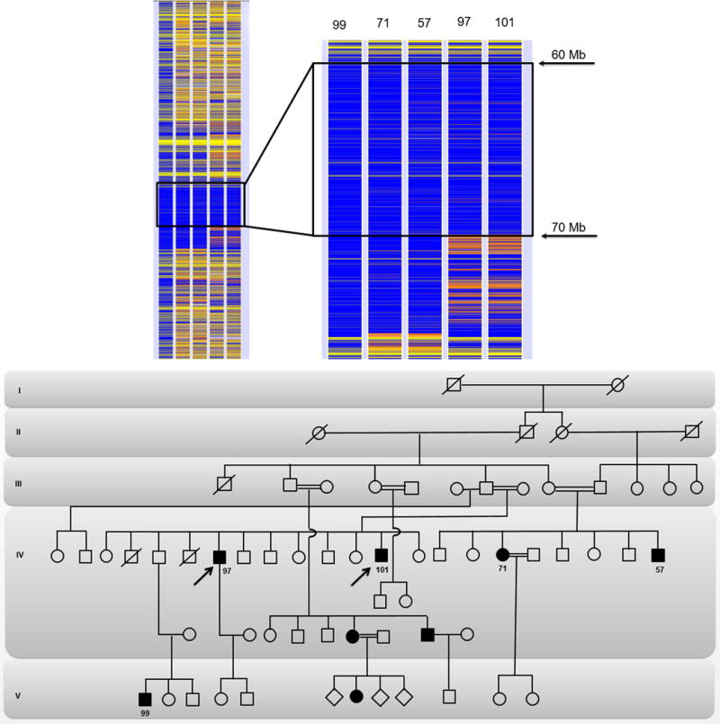
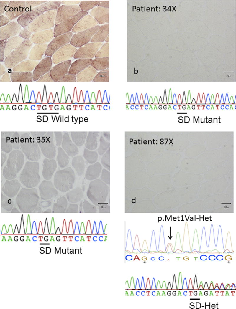
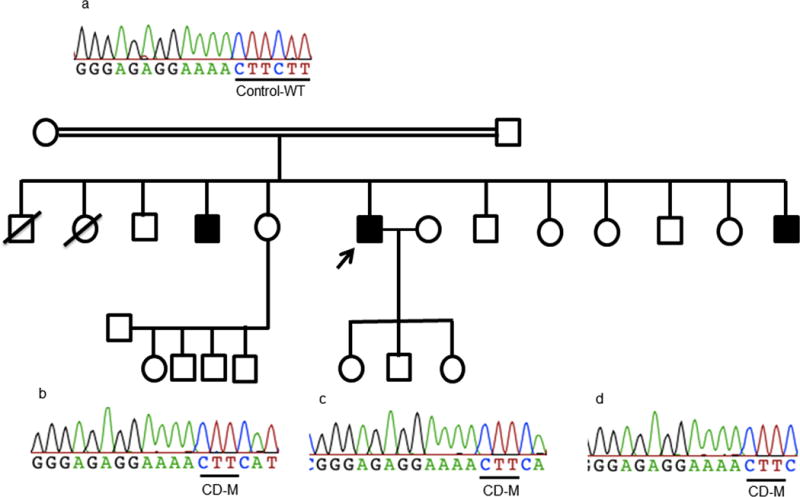
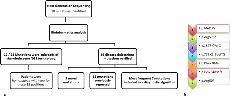
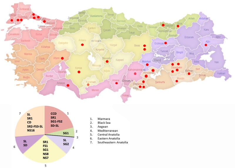
This study aimed to identify PYGM mutations in patients with McArdle disease from Turkey by next generation sequencing (NGS). Genomic DNA was extracted from the blood of the McArdle patients (n = 67) and unrelated healthy volunteers (n = 53). The PYGM gene was sequenced with NGS and the observed mutations were validated by direct Sanger sequencing. A diagnostic algorithm was developed for patients with suspected McArdle disease. A total of 16 deleterious PYGM mutations were identified, of which 5 were novel, including 1 splice-site donor, 1 frame-shift, and 3 non-synonymous variants. The p.Met1Val (27-patients/11-families) was the most common PYGM mutation, followed by p.Arg576* (6/4), c.1827+7A>G (5/4), c.772+2_3delTG (5/3), p.Phe710del (4/2), p.Lys754Asnfs (2/1), and p.Arg50* (1/1). A molecular diagnostic flowchart is proposed for the McArdle patients in Turkey, covering the 6 most common PYGM mutations found in Turkey as well as the most common mutation in Europe. The diagnostic algorithm may alleviate the need for muscle biopsies in 77.6% of future patients. A prevalence of any of the mutations to a geographical region in Turkey was not identified. Furthermore, the NGS approach to sequence the entire PYGM gene was successful in detecting a common missense mutation and discovering novel mutations in this population study.

Keywords: Genomics; Glycogenosis; Molecular screening; Novel mutation; Population specific; Rare muscle disorders.


Conflict of interest statement

Conflict of interest: None

Figures

Figure-1
Figure-1
Homozygosity map of chromosome 11 and zoom-in shared homozygote region by the affected individuals with p.Met1Val mutation is encircled in a black rectangle. The patient 99 in column 1 was arbitrarily selected as an index case. The variants of the index case are colored in blue and yellow, displaying homozygote and heterozygote areas, respectively. For other patients (columns 2 to 5), while a homozygote variant is indicated with blue, the contrasting homozygote variant is represented with white and the heterozygote variant with orange. For all patients, gray areas indicate no call and yellow indicate non-informative single nucleotide polymorphisms, which are heterozygote for the index case. The penetrance of the p.Met1Val mutation between generations of the large consanguineous family can be observed in the pedigree drawing while the numbers of individuals included in the homozygosity analysis are depicted below the circles and squares. The arrows represent the probands, the white circles and squares represent the non-myopathic subjects, the dark circles and squares represent the affected patients, and the ones with an oblique strait line represent the deceased family members.
Figure-2
Figure-2. Sequencing results of a consanguineous families with McArdle’s disease caused by NS8 and p.Arg576* (Stop gain 1-SG1)
The upper panel represents, a: heterozygote mother, b: heterozygote father, for NS8 and SG1 mutations respectively, c: the compound heterozygote affected son, and d: homozygote affected female relative only for the SG1 mutation. The nonsense mutation leads to premature termination of the myophosphorilase protein. The oblique arrows represent the probands in each family, the white circles and squares represent the non-myopathic subjects, the dark circles and squares represent the affected patients, and the ones with an oblique strait line represent the deceased family members. NS: non-synonymous, SG: stop gain, CD: codon deletion, Het: heterozygote, WT: wild type, M: mutant.
Figure-3
Figure-3. Pathology and sequencing results of patients homozygous and compound heterozygous for the novel c.772+2_3delTG splice site donor (SD) variant in PYGM gene
(a) Represents a wild type unrelated control pathology and sequence, (b) and (c) represents 2 unrelated homozygote mutant probands (patients 34 and 35), and (d) represents a third compound heterozgote proband (patient 87) for the novel SD mutation.
Figure-4
Figure-4
The p.Phe710del (codon deletion-CD) variants, the upper panel (a) represents a wild type unrelated control sequence, and (b), (c), and (d) represents 3 siblings homozygote for the p.Phe710del mutation. The arrows represent the probands in each family, the white circles and squares represent the non-myopathic subjects, the dark circles and squares represent the affected patients, and the ones with an oblique strait line represent the deceased family members. CD: codon deletion, WT: wild type, M: mutant.
Figure 5
Figure 5. Hierarchal task analysis and molecular flowchart for the diagnosis of McArdle disease in patients from Turkey
The NGS was successful in discovering 5 novel mutations in rare disorders, however also revealed the importance for cross-checking with the gold standard, the direct Sanger sequencing, as almost one third of the initially identified mutations by whole PYGM gene sequencing (12/28) were falsely called. Due to the high cost of NGS and the necessity to validate the mutations with direct sequencing a diagnostic flowchart was developed for future identification of patients with McArdle disease from Turkey. Based upon this proposed algorithm, 74.6% of patients (50/67) could be diagnosed molecularly including both homozygote and heterozygote patients for any of the mutations in the algorithm, and 58.2% of the patients (39/67) could be identified homozygous for 1 of the mutations in the algorithm. This algorithm is suggested for future patients from Turkey with suspected McArdle disease following a clinical examination.
Figure-6
Figure-6. The distribution of the families with McArdle disease who have volunteered to this study and a graphical analysis of the identified mutations in respect to the 7 geographical regions (1-7) of Turkey
Each red dot indicates 1 family, and represents their ancestral city of birth and the region. The pie analysis demonstrates the distribution of the mutations observed in this study population. The mutation of the 1 family that volunteered from the Aegean region (3) could not be identified hence the region could only be represented with the black line.

Similar articles

Cited by

References

    1. Darras BT, Friedman NR. Metabolic myopathies: a clinical approach; part II. Pediatr Neurol. 2000;22:171–81. - PubMed
    1. Chen YT. Glycogen storage diseases and other inherited disorders of carbohydrate metabolism. In: Kasper DL, Braunwald E, Fauci AS, Hauser SL, Longo DL, Jameson JL, editors. Harrison’s Principle of Internal Medicine. 16th. The McGraw-Hill Companies, Inc.; Pennsylvania, USA: 2005. pp. 2319–23.
    1. DiMauro S. Muscle glycogenoses: An overview. Acta Myol. 2007;26:35–41. - PMC - PubMed
    1. Lucia A, Nogales-Gadea G, Pérez M, Martín MA, Andreu AL, Arenas J. McArdle disease: what do neurologists need to know? Nat Clin Pract Neurol. 2008;4:568–77. - PubMed
    1. Deschauer M, Opalka JR, Lindner A, Zierz S. A novel nonsense mutation (R269X) in the myophosphorylase gene in a patient with McArdle disease. Mol Genet Metab. 2001;74:489–91. - PubMed

Substances