Clinical Trial
. 2017 Dec 6;25(12):2620-2634.
doi: 10.1016/j.ymthe.2017.08.016. Epub 2017 Aug 24.

Oncolytic H-1 Parvovirus Shows Safety and Signs of Immunogenic Activity in a First Phase I/IIa Glioblastoma Trial

Affiliations
Clinical Trial

Oncolytic H-1 Parvovirus Shows Safety and Signs of Immunogenic Activity in a First Phase I/IIa Glioblastoma Trial

Karsten Geletneky et al. Mol Ther. .

Abstract

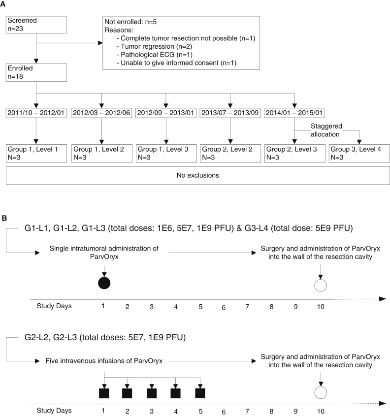
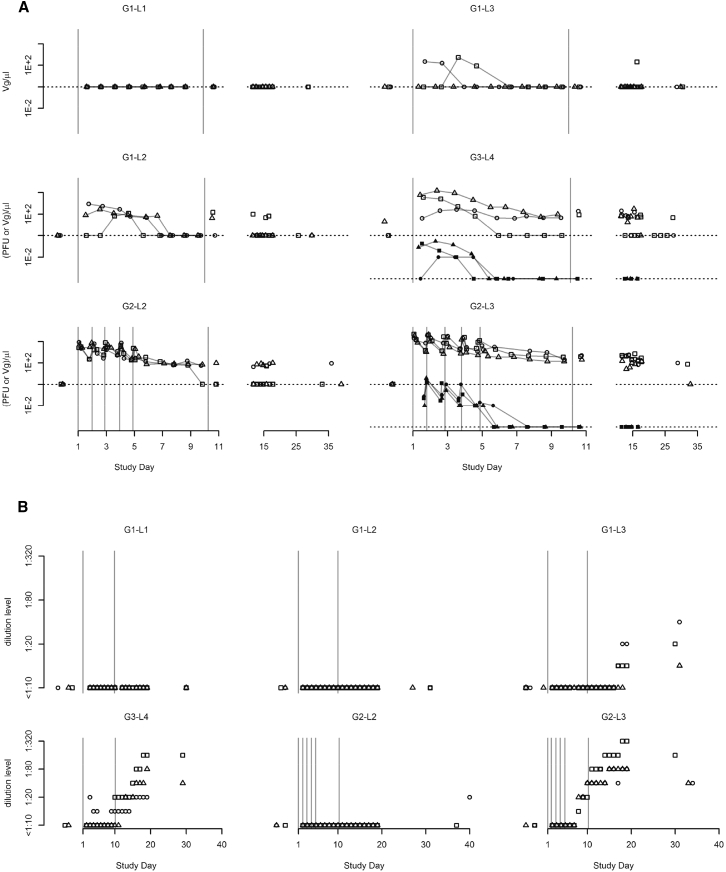
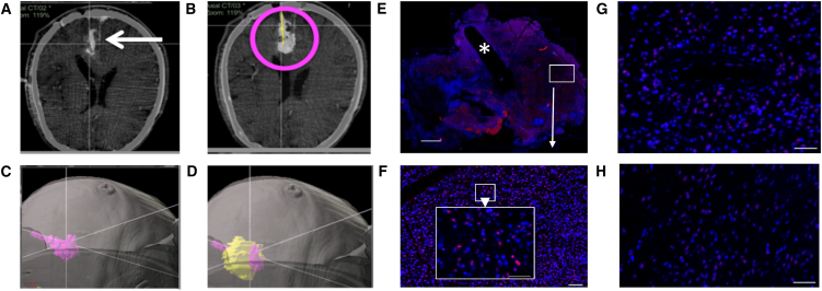
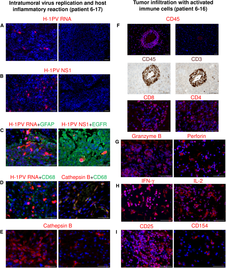
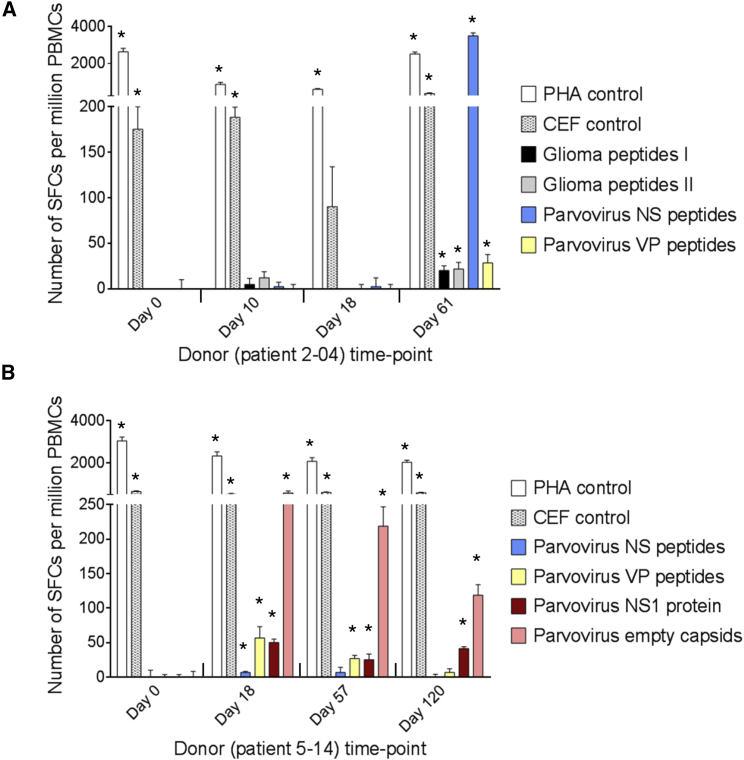
Oncolytic virotherapy may be a means of improving the dismal prognosis of malignant brain tumors. The rat H-1 parvovirus (H-1PV) suppresses tumors in preclinical glioma models, through both direct oncolysis and stimulation of anticancer immune responses. This was the basis of ParvOryx01, the first phase I/IIa clinical trial of an oncolytic parvovirus in recurrent glioblastoma patients. H-1PV (escalating dose) was administered via intratumoral or intravenous injection. Tumors were resected 9 days after treatment, and virus was re-administered around the resection cavity. Primary endpoints were safety and tolerability, virus distribution, and maximum tolerated dose (MTD). Progression-free and overall survival and levels of viral and immunological markers in the tumor and peripheral blood were also investigated. H-1PV treatment was safe and well tolerated, and no MTD was reached. The virus could cross the blood-brain/tumor barrier and spread widely through the tumor. It showed favorable pharmacokinetics, induced antibody formation in a dose-dependent manner, and triggered specific T cell responses. Markers of virus replication, microglia/macrophage activation, and cytotoxic T cell infiltration were detected in infected tumors, suggesting that H-1PV may trigger an immunogenic stimulus. Median survival was extended in comparison with recent meta-analyses. Altogether, ParvOryx01 results provide an impetus for further H-1PV clinical development.

Keywords: clinical trial; glioblastoma; oncolytic parvovirus; tumor microenvironment.


Figures

Figure 1
Figure 1
Schedule of ParvOryx Administration and Flow Chart of the Trial (A) Flow chart of the trial according to the CONSORT statement. The time interval assigned to each group and dose level represents the calendar period of patient enrollment into the corresponding cohort. (B) Schematic representation of the schedule of ParvOryx administration. Upper panel: treatment in G1 and G3. Intratumoral administration was performed through an intracranial catheter over approximately 30 min. Lower panel: treatment in G2. All five administrations were given as 2 hr intravenous infusions. In all groups on day 10, the remaining 50% of the total ParvOryx dose was injected into the walls of the resection cavity at multiple locations. PFU, plaque-forming units.
Figure 2
Figure 2
Pharmacokinetics and Seroconversion (A) Concentration over time, by cohort, of virus genomes (Vg; outline symbols) and infectious particles (PFU; solid symbols) in blood. Values below lower limits of quantification (LLOQ) are denoted by dotted lines. (B) Time course of anti-drug antibodies (ADAs) by cohort, as detected in a hemagglutination inhibition test.
Figure 3
Figure 3
Intratumoral Virus Distribution and Ability to Cross the Blood-Brain/Tumor Barrier (A–D) Distribution of the H-1PV inoculum after intratumoral injection (CT scan, patient 3-08). (A) Verification of correct catheter placement in a left occipital tumor by intraoperative CT prior to injection. (B) CT scan after injection of 1 mL of virus inoculum (magenta circle). (C) Three-dimensional segmenting of virus inoculum. (D) Overlay of reconstructed tumor (yellow) with virus inoculum (magenta), showing very little virus signal outside the tumor margins. (E and F) Virus distribution after intratumoral injection (patient 3-09). (E) FISH staining against H-1PV RNA of en bloc resected tumor with visible catheter track (asterisk). Scale bar, 2,000 μm. An area distant from the catheter track (white box) is magnified in (F) (white arrow). (F) Higher magnification (scale bar of whole image, 50 μm; scale bar of zoomed area, 100 μm) showing a strong hybridization signal for H-1PV RNA (red) at a distance of 7,000 μm from the catheter, thereby proving wide virus distribution through the tumor after local injection. (G and H) Intratumoral detection of H-1PV transcripts by FISH after intravenous injection (patient 4-10) indicating crossing of the blood-brain/tumor barrier. Hybridization signals are detected both around intratumoral blood vessels (G) and in blood vessel distant tumor areas (H). Scale bars, 50 μm.
Figure 4
Figure 4
In Situ Analysis of Tumors Resected after Local ParvOryx Administration (A–E) Intratumoral virus replication and host inflammatory reaction (patient 6-17). (A and B) H-1PV transcripts (A) and NS1 proteins (B) were detected in virus-injected tumor tissue (left), but not in historical controls (right). (C) Double staining was performed for (left) viral RNA (red) and glial fibrillary acidic protein (green), or (right) viral NS1 (red) and epidermal growth factor receptor (green). (D) H-1PV-transcript-accumulating tumor cells (red) stained negative for the macrophage marker CD68 (green) (left). In contrast, the majority of cathepsin B (CTSB)-positive cells (red) expressed CD68 (green) (right). CTSB+/CD68 cells were also detected (arrow). (E) Increased CTSB expression was observed in ParvOryx-treated tumor (left), as compared with historical control (right). (F–I) Tumor infiltration with activated immune cells (patient 6-16). (F) Upper two panels: the treated tumor showed increased leukocytic (CD45+) infiltration (left) compared with historical control (right). Middle two panels: tumor infiltrates (CD45, left) consisted predominantly of CD3+ T lymphocytes (right). Lower two panels: the T cell population included both CD8+ (left) and CD4+ (right) lymphocytes. (G–I) Several markers of immune cell activation were also detected in the ParvOryx-treated tumor: (G) granzyme B (left) and perforin (right), (H) IFN-γ (left) and IL-2 (right), and (I) CD25 (left) and CD154 (CD40L) (right). Scale bars, 50 μm.
Figure 5
Figure 5
Evaluation of T Cell Responses to H-1PV and Glioma Antigens by IFN-γ ELISpot Assay (A and B) Cellular immune responses are shown for two patients treated with ParvOryx via (A) the intratumoral and intracerebral route (patient 2-04) or (B) the intravenous and intracerebral route (patient 5-14). PBMCs were isolated at the indicated days prior to (day 0) or after (days 10–120) treatment. After incubation with appropriate stimulants, IFN-γ-producing spot-forming cells (SFCs) were counted. The test stimulants were viral or glioma peptides (Table S3) or full-length viral proteins (NS1 or empty capsids made of VP1 and VP2). Phytohemagglutinin (PHA) and cytomegalovirus, Epstein-Barr virus, and influenza virus (CEF) peptide pools served as positive control stimulants. Negative control values (unstimulated cells) ranged from 0 to 21 SFCs per million PBMCs and were subtracted from the corresponding stimulated sample values. Means (columns) and SEMs (bars) of triplicate measurements are shown. Asterisks denote statistical significance (*p ≤ 0.05; mean SFC − 2 SEMs > 2× negative control).

References

    1. Stupp R., Hegi M.E., Mason W.P., van den Bent M.J., Taphoorn M.J., Janzer R.C., Ludwin S.K., Allgeier A., Fisher B., Belanger K., European Organisation for Research and Treatment of Cancer Brain Tumour and Radiation Oncology Groups. National Cancer Institute of Canada Clinical Trials Group Effects of radiotherapy with concomitant and adjuvant temozolomide versus radiotherapy alone on survival in glioblastoma in a randomised phase III study: 5-year analysis of the EORTC-NCIC trial. Lancet Oncol. 2009;10:459–466. - PubMed
    1. Clarke J.L., Ennis M.M., Yung W.K., Chang S.M., Wen P.Y., Cloughesy T.F., Deangelis L.M., Robins H.I., Lieberman F.S., Fine H.A., North American Brain Tumor Consortium Is surgery at progression a prognostic marker for improved 6-month progression-free survival or overall survival for patients with recurrent glioblastoma? Neuro-oncol. 2011;13:1118–1124. - PMC - PubMed
    1. Russell S.J., Peng K.-W., Bell J.C. Oncolytic virotherapy. Nat. Biotechnol. 2012;30:658–670. - PMC - PubMed
    1. Miest T.S., Cattaneo R. New viruses for cancer therapy: meeting clinical needs. Nat. Rev. Microbiol. 2014;12:23–34. - PMC - PubMed
    1. Lichty B.D., Breitbach C.J., Stojdl D.F., Bell J.C. Going viral with cancer immunotherapy. Nat. Rev. Cancer. 2014;14:559–567. - PubMed

Publication types

MeSH terms