Review
. 2017 Oct 24;56(42):5628-5634.
doi: 10.1021/acs.biochem.7b00747. Epub 2017 Oct 10.

Structural Basis for G Protein-Coupled Receptor Activation

Affiliations
Review

Structural Basis for G Protein-Coupled Receptor Activation

Aashish Manglik et al. Biochemistry. .

Abstract

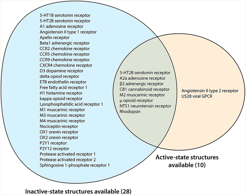
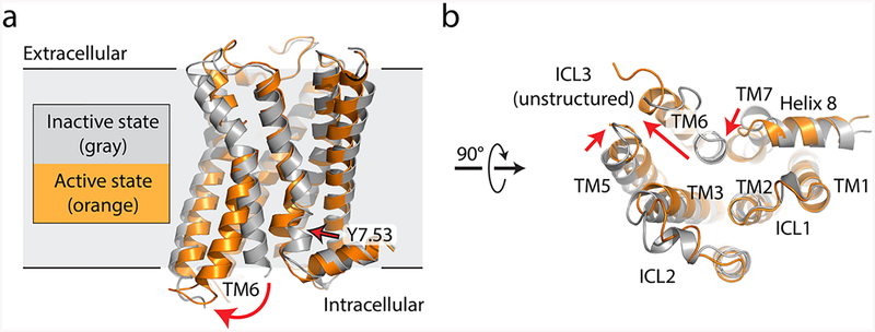
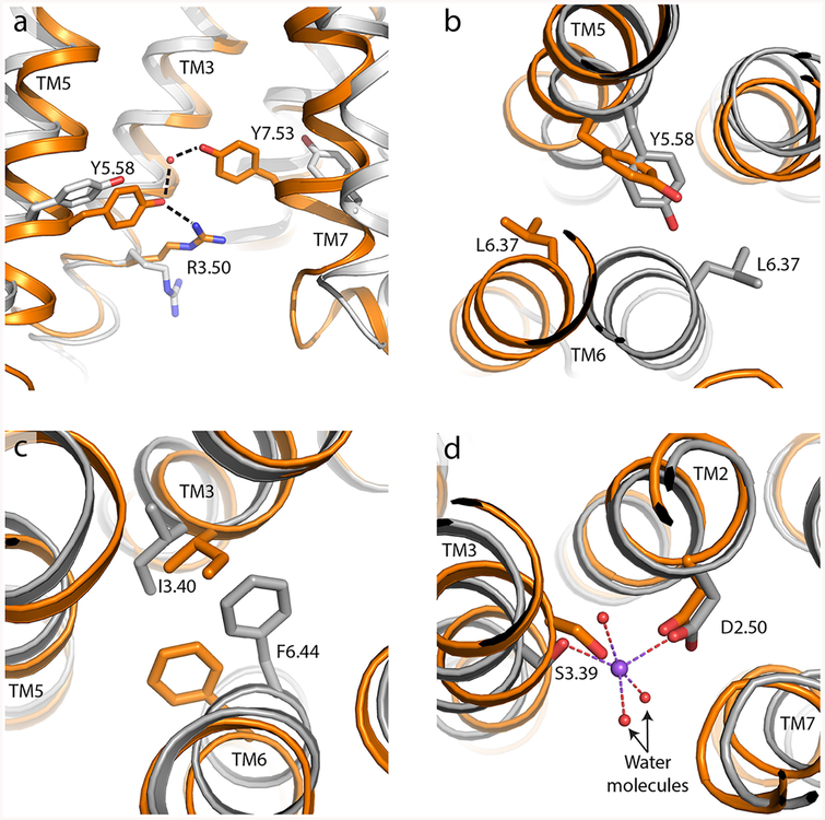
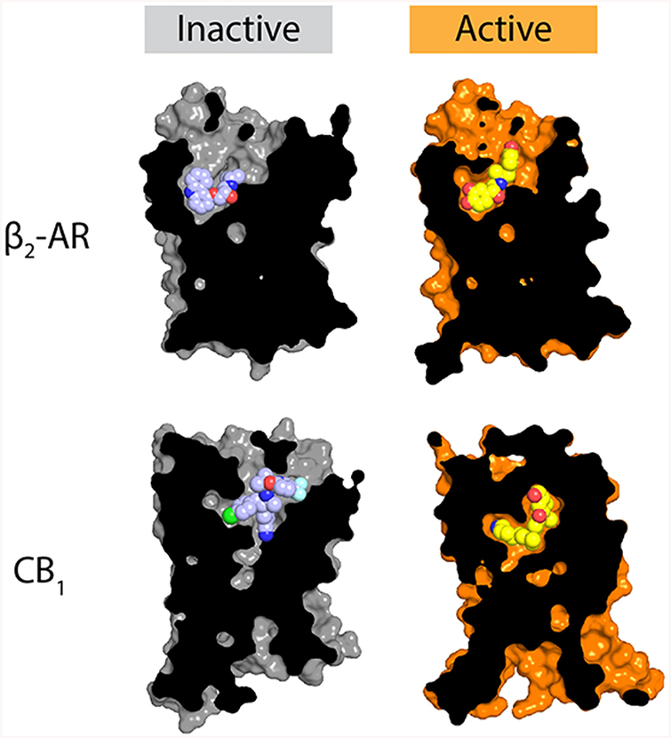
G protein-coupled receptors (GPCRs) are critical regulators of human physiology and make up the largest single class of therapeutic drug targets. Although GPCRs regulate highly diverse physiology, they share a common signaling mechanism whereby extracellular stimuli induce conformational changes in the receptor that enable activation of heterotrimeric G proteins and other intracellular effectors. Advances in GPCR structural biology have made it possible to examine ligand-induced GPCR activation at an unprecedented level of detail. Here, we review the structural basis for family A GPCR activation, with a focus on GPCRs for which structures are available in both active or active-like states and inactive states. Crystallographic and other biophysical data show how chemically diverse ligands stabilize highly conserved conformational changes on the intracellular side of the receptors, allowing many different extracellular stimuli to utilize shared downstream signaling molecules. Finally, we discuss the remaining challenges in understanding GPCR activation and signaling and highlight new technologies that may allow unanswered questions to be resolved.


Conflict of interest statement

Notes

The authors declare no competing financial interest.

Figures

Figure 1.
Figure 1.
Family A GPCRs for which structural data are available. Most family A GPCRs that have been crystallized to date have been determined in only a single conformational state, usually an inactive conformation.
Figure 2.
Figure 2.
Comparison of active and inactive states for a prototypical GPCR. (a) Inactive-state (gray, PDB entry 3UON) and active-state (orange, PDB entry 4MQS) structures of the human M2 muscarinic acetylcholine receptor are shown in a side view, parallel to the membrane plane. Red arrows indicate conformational changes upon activation. (b) Same structure, viewed from the intracellular side.
Figure 3.
Figure 3.
Intracellular motifs involved in GPCR activation. In each case, the inactive receptor is colored gray and the active state of the same receptor is colored orange. (a) Side view showing Tyr5.58 and Tyr7.53 engaged in a highly conserved hydrogen bond mediated by a bridging water molecule (red sphere), often also interacting with Arg3.50 as seen here for the μ-opioid receptor (PDB entries 4DKL for the inactive state and 5C1M for the active state). (b) Leucine ratchet of Leu6.37 past Tyr5.58 upon activation, exemplified by the β2 adrenergic receptor viewed from the extracellular direction (PDB entries 2RH1 for the inactive state and 4LDE for the active state). (c) Phe-Tyr switch in the β2 adrenergic receptor (same PDB entries as in panel b, also viewed from above/extracellular). (d) Structural sodium ion stabilization of the inactive-state receptor, shown here for the A2a adenosine receptor viewed from above (PDB entries 4EIY for the inactive state and 5G53 for the active state).
Figure 4.
Figure 4.
Agonist recognition. The molecular details of agonist recognition are highly diverse, although most agonist-bound activate-state GPCR structures show a modest contraction of the ligand binding site relative to their inactive-state counterparts. Here, the structures of the inactive and active β2 adrenergic receptor (PDB entries 2RH1 and 4LDL, respectively) and the CB1 cannabinoid receptor (PDB entries 5U09 and 5XRA for the inactive and active states, respectively) are shown as representative examples, showing contraction of the binding site upon activation, as well as the far more extensive nature of structural rearrangements upon activation of the CB1 receptor compared to the β2 receptor.

References

    1. Rosenbaum DM, Cherezov V, Hanson MA, Rasmussen SG, Thian FS, Kobilka TS, Choi HJ, Yao XJ, Weis WI, Stevens RC, and Kobilka BK (2007) GPCR engineering yields high-resolution structural insights into beta2-adrenergic receptor function. Science 318, 1266–1273. - PubMed
    1. Chae PS, Rasmussen SG, Rana RR, Gotfryd K, Chandra R, Goren MA, Kruse AC, Nurva S, Loland CJ, Pierre Y, Drew D, Popot JL, Picot D, Fox BG, Guan L, Gether U, Byrne B, Kobilka B, and Gellman SH (2010) Maltose-neopentyl glycol (MNG) amphiphiles for solubilization, stabilization and crystallization of membrane proteins. Nat. Methods 7, 1003–1008. - PMC - PubMed
    1. Caffrey M (2015) A comprehensive review of the lipid cubic phase or in meso method for crystallizing membrane and soluble proteins and complexes. Acta Crystallogr., Sect. F: Struct. Biol. Commun 71, 3–18. - PMC - PubMed
    1. Smith JL, Fischetti RF, and Yamamoto M (2012) Micro-crystallography comes of age. Curr. Opin. Struct. Biol 22, 602–612. - PMC - PubMed
    1. Johansson LC, Stauch B, Ishchenko A, and Cherezov V (2017) A Bright Future for Serial Femtosecond Crystallography with XFELs. Trends Biochem. Sci 42, 749–762. - PMC - PubMed

Publication types

MeSH terms

Substances