. 2017 Dec;18(12):1299-1309.
doi: 10.1038/ni.3853. Epub 2017 Oct 2.

NLRX1 promotes immediate IRF1-directed antiviral responses by limiting dsRNA-activated translational inhibition mediated by PKR

Affiliations

NLRX1 promotes immediate IRF1-directed antiviral responses by limiting dsRNA-activated translational inhibition mediated by PKR

Hui Feng et al. Nat Immunol. 2017 Dec.

Abstract

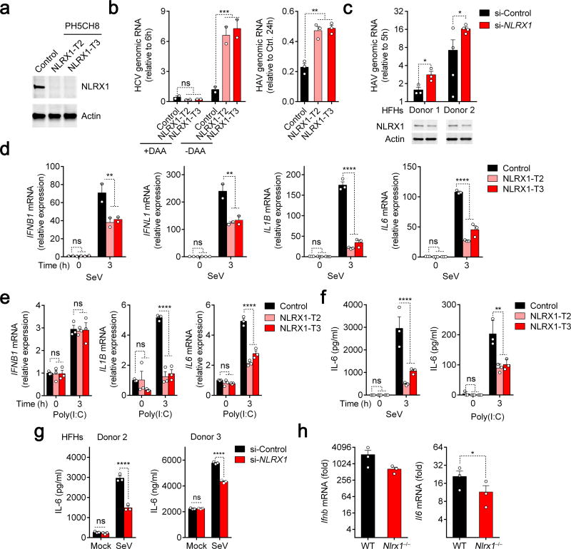
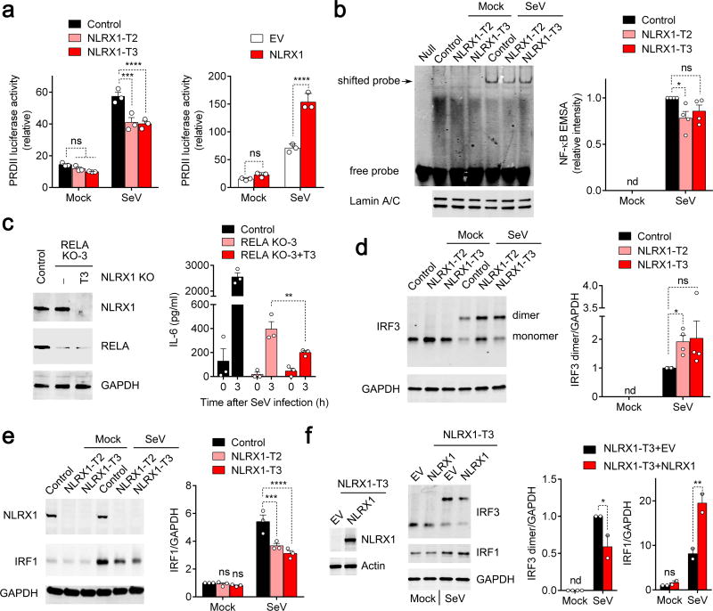
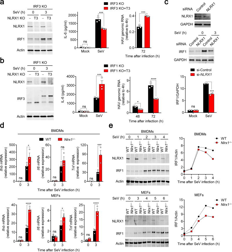
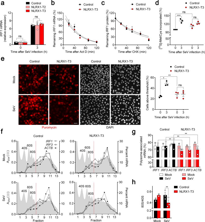
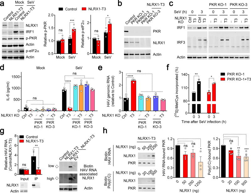
NLRX1 is unique among the nucleotide-binding-domain and leucine-rich-repeat (NLR) proteins in its mitochondrial localization and ability to negatively regulate antiviral innate immunity dependent on the adaptors MAVS and STING. However, some studies have suggested a positive regulatory role for NLRX1 in inducing antiviral responses. We found that NLRX1 exerted opposing regulatory effects on viral activation of the transcription factors IRF1 and IRF3, which might potentially explain such contradictory results. Whereas NLRX1 suppressed MAVS-mediated activation of IRF3, it conversely facilitated virus-induced increases in IRF1 expression and thereby enhanced control of viral infection. NLRX1 had a minimal effect on the transcription of IRF1 mediated by the transcription factor NF-kB and regulated the abundance of IRF1 post-transcriptionally by preventing translational shutdown mediated by the double-stranded RNA (dsRNA)-activated kinase PKR and thereby allowed virus-induced increases in the abundance of IRF1 protein.


Conflict of interest statement

COMPETING FINANCIAL INTERESTS

The authors declare no competing financial interests.

Figures

Figure 1
Figure 1
NLRX1 is a positive regulator of innate immunity and an antiviral factor in hepatocytes. (a) Immunoblot confirmation of NLRX1 depletion in two independent PH5CH8-derived cell lines. (b) qRT-PCR of HCV and HAV genomic RNAs in NLRX1-deficient cells transfected with HCV JFH1-QL RNA (left, 24 h) or infected with HAV (right, 72 h). Data are from n=2 (left) or 3 (right) technical replicates and are representative of 3 independent experiments. (c) qRT-PCR of HAV genomic RNA in primary human fetal hepatoblasts (HFHs) with partial NLXR1 depletion at 72 h. n=3 and 4 technical replicates for donors 1 and 2, respectively. (d,e) qRT-PCR for IFNB1, IFNL1, IL1B and IL6 mRNA in NLRX1-deficient PH5CH8 cells infected with SeV (d) or stimulated with poly(I:C) added to medium for 3 h (e). Data are from 2 independent experiments with 4 technical replicates (d, IFNB1 and IFNL1), or are representative of 3 experiments, each with n=3 technical replicates (d, IL1B and IL6; e). (f,g) ELISA for IL-6 protein production in NLRX1-deficient PH5CH8 cells (f) and NLRX1-depleted HFHs (g). Data are representative of 3 experiments or donors, each with n=3 technical replicates. (h) qRT-PCR for Ifnb and Il6 mRNA in livers of Nlrx1−/− mice 3 h following hydrodynamic injection of HAV RNA (n=3 per group). For all qRT-PCR analyses, host gene expression was normalized to ACTB (human) or Actb (mouse). All data are shown as mean ± S.E.M. Unless otherwise indicated, comparisons were between control and NLRX1-depleted cells by two-way ANOVA (ns, not significant; *p < 0.05; ** p< 0.01; ***p < 0.001; ****p < 0.0001). Comparisons in (b right,c,g,h) were performed by t test (*p < 0.05).
Figure 2
Figure 2
Impact of NLRX1 deficiency on NF-κB signaling and IRF1 and IRF3 activation in SeV-infected PH5CH8 cells. (a) PRDII-Luc promoter activation in cells with NLRX1 depletion (left) or overexpression (right). Data are from n=3 technical replicates and are representative of 3 independent experiments. (b) NF-κB electrophoretic mobility shift assay (EMSA) with nuclear extracts from mock- and SeV-infected NLRX1-deficient PH5CH8 cells (left). Mean infrared fluoresence intensity measurements from 4 independent EMSA experiments (right). (c) Effects of NLRX1 depletion in the absence of RELA. Immunoblots for NLRX1 and RELA in NLRX1-RELA double-deficient (RELA KO+NLRX1-T3) PH5CH8 cells (left). ELISA quantitation of IL-6 protein abundance in SeV-infected (3 h) control, RELA-deficient and NLRX1-RELA double-deficient cells (right). Results are representative of 2 independent experiments, each with 3 technical repeats. (d,e) Immunoblots showing SeV-triggered IRF3 dimerization (d) and IRF1 expression in NLRX1-deficient cells (e). n=3–4 independent experiments. (f) SeV-induced IRF3 dimerization and increased IRF1 protein expression in NLRX1-reconstituted NLRX1-T3 cells. Data are representative of 2 independent experiments. All data are shown as mean ± S.E.M. Comparisons of control versus NLRX1-depleted cells in panels (a,e) and (b-d, f) were made by two-way ANOVA and t test, respectively. ns = nonsignificant, *p< 0.05; **p < 0.01; ***p< 0.001; ****p< 0.0001.
Figure 3
Figure 3
NLRX1/IRF1 signaling dominates the antiviral cytokine response in hepatocytes. (a,b) ELISA measurements of SeV-induced IL-6 production and qRT-PCR quantitation of HAV replication in IRF3-deficient and IRF3-NLRX1 double-deficient PH5CH8 cells (a), or IRF1-deficient and IRF1-NLRX1 double-deficient cells (b). Panels on the left show immunoblots for NLRX1 and IRF1 (a) or IRF3 dimerization at 3 h (b). See Supplementary Fig. 2f for immunoblots of IRF1-deficient and IRF3-deficient cells. Results are representative of 2–3 independent experiments, each with 3 technical repeats. (c) Immunoblots of NLRX1 and IRF1 in partial NLRX1-depleted HFHs infected with SeV for 3 h. Bottom panel shows quantification of immunoblots (infrared fluorescence intensities) from 2 independent experiments using cells from different donors. (d) qRT-PCR for Ifnb, Il6 and Tnf mRNA and (e) immunoblots of IRF1 in SeV-infected wild-type versus Nlrx1−/− BMDMs (top panels) and MEFs (bottom panels). Data are from n=3 technical replicates and are representative of 4 (BMDMs) and 2 (MEFs) independent experiments. All immunoblots are representative of 2–3 independent experiments or donors. Data are shown as mean ± S.E.M. ns = nonsignificant, *p< 0.05; **p < 0.01; ***p< 0.001; ****p< 0.0001 by t test.
Figure 4
Figure 4
NLRX1 facilitates immediate IRF1 antiviral responses by promoting global protein synthesis in SeV-infected cells. (a) qRT-PCR of SeV-induced IRF1 mRNA responses in control and NLRX1-deficient PH5CH8 cells. (n=3). (b) IRF1 mRNA stability in actinomycin D (Act D)-treated SeV-infected control vs. NLRX1-T3 PH5CH8 cells. (n=3). (c) IRF1 protein stability in cycloheximide (CHX)-treated cells following SeV infection assessed by quantitation of immunoblots. (n=6). See Supplementary Fig. 4b for representative immunoblots. (d) Nascent protein synthesis monitored by [35S]-Met/Cys incorporation in mock- versus SeV-infected control and NLRX1-T3 cells. [35S] incorporated into TCA-precipitated protein is shown relative to that in uninfected control cells (100%). Data are from n=6 technical replicates from three experiments. See Supplementary Fig. 4d for representative immunoblots. (e) Global protein synthesis imaged by confocal microscopy in mock- versus SeV-infected control and NLRX1-T3 cells. Puromycin was detected by staining with specific antibody, and nuclei counterstained with DAPI. Scale bar, 40 µm. The percentage of cells with puromycin labeling exceeding an arbitrary threshold in 3 experiments is shown on the right. An average of 176 cells were evaluated for each condition. (f) Representative polysome profiles of IRF1, IRF3 and ACTB mRNAs in mock- versus SeV-infected control and NLRX1-T3 cells. Absorbance at 254 nm (A254) is aligned with qRT-PCR quantitation of mRNA, plotted as percent of total mRNA in each fraction. (g) Proportion of IRF1, IRF3 and ACTB mRNAs associated with translationally-active polysomes (fractions 7–14) in SeV- (n=3) and mock-infected (n=2) control versus NLRX1-T3 cells. (h) 80S/40S ratios determined from the area under the curve (AUC) of 40S and 80S peaks of A254 traces in 2–3 experiments. Data are shown as mean ± S.E.M. ns = nonsignificant, *p< 0.05; **p < 0.01; ***p< 0.001 by two-way ANOVA (a,d,e,g) or t test (h).
Figure 5
Figure 5
NLRX1 reduces PKR activation and subsequent inhibition of protein synthesis by competing for viral RNA binding. (a) Immunoblots of PKR-eIF2α signaling in NLRX1-deficient PH5CH8 cells 4.5 h after SeV infection. Quantification (infrared fluorescence intensity) of p-PKR or p-eIF2α was normalized to actin (n=4 experiments). (b) Immunoblots of PKR and NLRX1 in PKR-deficient and NLRX1-PKR double-deficient PH5CH8 cells. (c) Immunoblots of SeV-induced IRF1 expression and IRF3 dimerization in NLRX1-deficient and NLRX1-PKR double-deficient PH5CH8 cells. The image was generated from 2 separate gels in one experiment; data are representative of 2 experiments. (d,e) ELISA for SeV-triggered IL-6 protein production (d) and qRT-PCR for HAV RNA (e) in control, NLRX1-deficient and NLRX1-PKR double-deficient PH5CH8 cells. Data are representative of 2–3 experiments, each with 3 technical replicates. (f) [35S]-Met/Cys incorporation as measurement of global protein synthesis in SeV- versus mock-infected NLRX1-PKR double-deficient PH5CH8 cells. Data are from 4 technical replicates of 2 experiments. (g) Bidirectional assessment of NLRX1-HAV RNA association. (left) qRT-PCR of HAV RNA co-immunoprecipitating with NLRX1. HAV RNA was transfected as in Fig. 1b, followed by immunoprecipitation with anti-NLRX1 at 3 h. Immunoblots of input and immunoprecipitated (IP) NLRX1 are shown at the bottom. Data are representative of 2 experiments (n=2–3). (right) Immunoblots of NLRX1 co-precipitating with biotin-tagged HAV RNA in cell lysates. Immunoblots are representative of 3 experiments (◇ indicates endogenous NLRX1 associated with HAV RNA). “High” and “Low” indicate detection intensity. (h) Impact of NLRX1 reconstitution on PKR association with biotin-tagged HAV RNA or poly(I:C) in NLRX1-T3 cell lysates. Immunoblots are representative of 3 (HAV) or 5 [poly(I:C)] experiments. Quantification of PKR binding is summarized on the right. Data are shown as mean ± S.E.M. ns = nonsignificant, *p< 0.05; **p < 0.01; ***p< 0.001 by two-way ANOVA (a,d,f,g) and one-way ANOVA (e,h).

References

    1. Guo H, Callaway JB, Ting JP. Inflammasomes: mechanism of action, role in disease, and therapeutics. Nat Med. 2015;21:677–687. - PMC - PubMed
    1. Schneider M, et al. The innate immune sensor NLRC3 attenuates Toll-like receptor signaling via modification of the signaling adaptor TRAF6 and transcription factor NF-kappaB. Nat Immunol. 2012;13:823–831. - PMC - PubMed
    1. Xia X, et al. NLRX1 negatively regulates TLR-induced NF-kappaB signaling by targeting TRAF6 and IKK. Immunity. 2011;34:843–853. - PMC - PubMed
    1. Benko S, Magalhaes JG, Philpott DJ, Girardin SE. NLRC5 limits the activation of inflammatory pathways. J Immunol. 2010;185:1681–1691. - PubMed
    1. Allen IC, et al. NLRP12 suppresses colon inflammation and tumorigenesis through the negative regulation of noncanonical NF-kappaB signaling. Immunity. 2012;36:742–754. - PMC - PubMed

MeSH terms