. 2017 Oct 2;12(10):e0185664.
doi: 10.1371/journal.pone.0185664. eCollection 2017.

Preferential activation of HIF-2α adaptive signalling in neuronal-like cells in response to acute hypoxia

Affiliations

Preferential activation of HIF-2α adaptive signalling in neuronal-like cells in response to acute hypoxia

Miguel A S Martín-Aragón Baudel et al. PLoS One. .

Abstract

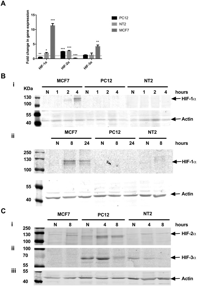
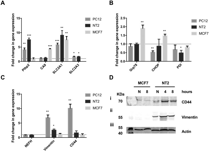
Stroke causes severe neuronal damage as disrupted cerebral blood flow starves neurons of oxygen and glucose. The hypoxia inducible factors (HIF-1α and HIF-2α) orchestrate oxygen homeostasis and regulate specific aspects of hypoxic adaptation. Here we show the importance of HIF-2α dependant signalling in neuronal adaptation to hypoxic insult. PC12 and NT2 cells were differentiated into neuronal-like cells using NGF and retinoic acid, and exposed to acute hypoxia (1% O2). Gene and protein expression was analysed by qPCR and immunoblotting and the neuronal-like phenotype was examined. PC12 and NT2 differentiation promoted neurite extension and expression of neuronal markers, NSE and KCC2. Induction of HIF-1α mRNA or protein was not detected in hypoxic neuronal-like cells, however marked induction of HIF-2α mRNA and protein expression was observed. Induction of HIF-1α target genes was also not detected in response to acute hypoxia, however significant induction of HIF-2α transcriptional targets was clearly evident. Furthermore, hypoxic insult dramatically reduced both neurite number and length, and attenuated expression of neuronal markers, NSE and KCC2. This correlated with an increase in expression of the neural progenitor and stem cell-like markers, CD44 and vimentin, suggesting HIF-2α molecular mechanisms could potentially promote regression of neuronal-like cells to a stem-like state and trigger neuronal recovery following ischaemic insult. Our findings suggest the HIF-2α pathway predominates over HIF-1α signalling in neuronal-like cells following acute hypoxia.


Conflict of interest statement

Competing Interests: The authors have declared that no competing interests exist.

Figures

Fig 1
Fig 1. Characterising PC12 and NT2 morphology, gene and protein expression following differentiation into neuronal-like cells.
A: Representative bright-field microscopy images of PC12 cell morphology after 3 and 8 days in the presence of NGF, (20x magnification). B: Relative Nrn1, Tmod1 and Caspr1 mRNA expression in differentiated PC12 cells was analysed by qPCR. C: Representative bright-field microscopy images of NT2 cell morphology were taken 4 weeks after treatment with all-trans-retinoic acid (ATRA 4w) and a further 2 weeks after treatment with cytosine arabinoside (Ara-C) (ATRA 4w + 2w Ara-C); (20x magnification). D: Relative Nrn1, Tmod1 and Caspr1 mRNA expression in differentiated NT2 cells was analysed by qPCR. E: Representative immunoblot analysis showing NSE and KCC2 protein expression in undifferentiated (U) and differentiated (D) PC12 and NT2 cells. Equal protein loading was assessed by immunoblotting for actin. A and C: Scale bar represents 50 μm; white arrows indicate interlaced axon-like structures and black arrows indicate ganglion-like clusters. B and D: Data is presented as the mean ± SEM; n = 3; **p≤0.01, ***p≤0.001.
Fig 2
Fig 2. The effect of hypoxia on neuronal-like PC12 and NT2 cells.
A: MCF7, PC12 and NT2 mitochondrial activity was analysed using alamarBlue 2, 4, 8 and 24 hours after exposure to hypoxia and expressed as a percentage of normoxic cell activity. B: MCF7, PC12 and NT2 cell viability was analysed via trypan blue staining 8 hours after exposure to hypoxia (8) and compared to staining of normoxic (N) cells. Data is expressed as mean ± SEM; n = 3; ***p≤0.001.
Fig 3
Fig 3. Increased HIF-2α stability is observed in neuronal-like PC12 and NT2 cells following hypoxia.
A: Relative HIF1-3α mRNA expression was analysed in MCF7 and differentiated PC12 and NT2 cells exposed to 8 hours of hypoxia by qPCR. Data is expressed as mean ± SEM; n = 3; *p<0.05, **p≤0.01, ***p≤0.001. The dotted line represents basal gene expression. B-C: Representative immunoblots of HIF-1α (B), HIF-2α (Ci), HIF-3α (Cii) protein expression in MCF7 and differentiated PC12 and NT2 cells, 1, 2, 4, 8 or 24 hours after exposure to hypoxia. B and C: Equal protein loading was assessed by immunoblotting for actin.
Fig 4
Fig 4. HIF mediated adaptation to hypoxic stress.
A: Under physiological oxygen concentrations, HIF-1α/2α are hydroxylated by prolyl hydroxylases (PHD), promoting HIF-1α/2α binding to the E3 ligase, von Hippel-Lindau protein (pVHL), and their ubiquitination [3]. This maintains very low basal expression of HIF-1α/2α due to rapid proteasomal degradation. B: Under hypoxic conditions, PHD activity is inhibited [38]. This stabilises HIF-1α/2α expression, enhancing binding to HIFβ and translocation to the nucleus and transcription of various hypoxia-responsive genes[3]. HIF-1α and -2α share regulation of several genes, yet also regulate distinct subsets [4,6]. HIF-3α function is not yet fully understood. Abbreviations: CHOP, CCAAT-enhancer-binding protein homologous protein; EPO, Erythropoietin; Grp, Glucose regulated protein; HIF: Hypoxia Inducible Factor; PDI, protein disulphide isomerase; PHD, Prolyl hydroxylase; VEGF, Vascular epithelial growth factor; VHL, Von Hippel Lindau.
Fig 5
Fig 5. HIF-2α dependant pathways are preferentially activated in differentiated PC12 and NT2 cells following hypoxia.
Relative expression of HIF-1α related target genes (Ptbp2, CA9, SLC2A1 and SLC2A3) (A); UPR related genes (Grp78, CHOP and PDI) (B); and HIF-2α related target genes (NEFH, Vimentin and CD44) (C), were analysed in MCF7 and differentiated PC12 and NT2 cells, 8 hours after hypoxia, using qPCR. Data is expressed as mean ± SEM; n = 3; *p<0.05, **p≤0.01, ***p≤0.001. The dotted line represents basal gene expression. D: Representative immunoblots showing induction of CD44 (i) and vimentin (ii) protein expression in normoxic (N) or hypoxic (4 or 8 hours hypoxia) MCF7 and differentiated NT2 cells. Equal protein loading was assessed by immunoblotting for actin (iii).
Fig 6
Fig 6. Hypoxia promotes a regression of neuronal-like cells to undifferentiated states.
A: Representative immunoblots showing the change in NSE (i) and KCC2 (ii) protein expression in normoxic (N), differentiated (D) or differentiated and hypoxic (D+H, 8 hours) PC12 and NT2 cells. Equal protein loading was assessed by immunoblotting for actin (iii). B: Representative bright-field microscopy images of differentiated PC12 and NT2 morphology after exposure to 8 hours of normoxia or hypoxia (20x magnification). Scale bar represents 50 μm. White arrows indicate interlaced axon-like structures and black arrows indicate ganglion-like clusters.

References

    1. Astrup J, Siesjö BK, Symon L. Thresholds in cerebral ischemia—the ischemic penumbra. Stroke. 12: 723–5. Available: http://www.ncbi.nlm.nih.gov/pubmed/6272455 - PubMed
    1. Liu S, Levine SR, Winn HR. Targeting ischemic penumbra: part I—from pathophysiology to therapeutic strategy. J Exp Stroke Transl Med. NIH Public Access; 2010;3: 47–55. - PMC - PubMed
    1. Semenza GL. Hypoxia-inducible factors in physiology and medicine. Cell. 2012. pp. 399–408. doi: 10.1016/j.cell.2012.01.021 - DOI - PMC - PubMed
    1. Hu C-J, Wang L-Y, Chodosh LA, Keith B, Simon MC. Differential roles of hypoxia-inducible factor 1alpha (HIF-1alpha) and HIF-2alpha in hypoxic gene regulation. Mol Cell Biol. 2003;23: 9361–74. Available: http://www.ncbi.nlm.nih.gov/pubmed/14645546 doi: 10.1128/MCB.23.24.9361-9374.2003 - DOI - PMC - PubMed
    1. Keith B, Johnson RS, Simon MC. HIF1α and HIF2α: sibling rivalry in hypoxic tumour growth and progression. Nat Rev Cancer. 2012;12: 9–22. doi: 10.1038/nrc3183 - DOI - PMC - PubMed

Substances