Review
. 2018 May 10;61(9):3755-3778.
doi: 10.1021/acs.jmedchem.7b00574. Epub 2017 Oct 19.

Modulators of 14-3-3 Protein-Protein Interactions

Affiliations
Review

Modulators of 14-3-3 Protein-Protein Interactions

Loes M Stevers et al. J Med Chem. .

Abstract

Direct interactions between proteins are essential for the regulation of their functions in biological pathways. Targeting the complex network of protein-protein interactions (PPIs) has now been widely recognized as an attractive means to therapeutically intervene in disease states. Even though this is a challenging endeavor and PPIs have long been regarded as "undruggable" targets, the last two decades have seen an increasing number of successful examples of PPI modulators, resulting in growing interest in this field. PPI modulation requires novel approaches and the integrated efforts of multiple disciplines to be a fruitful strategy. This perspective focuses on the hub-protein 14-3-3, which has several hundred identified protein interaction partners, and is therefore involved in a wide range of cellular processes and diseases. Here, we aim to provide an integrated overview of the approaches explored for the modulation of 14-3-3 PPIs and review the examples resulting from these efforts in both inhibiting and stabilizing specific 14-3-3 protein complexes by small molecules, peptide mimetics, and natural products.


Conflict of interest statement

The authors declare no competing financial interest.

Figures

Figure 1
Figure 1
Schematic depicting different strategies for modulation of PPIs: competitive (orthosteric) inhibition (A), allosteric inhibition (B), and stabilization (C).
Figure 2
Figure 2
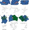
Representative examples of competitive, allosteric, and stabilizing PPI ligands (X-ray structure (above), chemical structure (below)). (A) p53/hDM2 inhibitor RG7112 (PDB ID: 4IPF). (B) BH3/Bcl-2 inhibitor ABT-199 (PDB ID: 4MAN). (C) Fragment of BRD4/Histone inhibitor I-BET762 (PDB ID: 4C66). (D) HIF-2 PAS domain allosteric modulator (PDB ID: 4GHI). (E) FKBP12/Rapamycin/FRAP stabilizer complex (PDB ID: 1FAP). (F) Transthyretin stabilizer (PDB ID: 2FLM).
Figure 3
Figure 3
14-3-3 structure and binding of partner protein peptides exemplified by the 14-3-3ζ/C-Raf complex (PDB ID: 4FJ3). Top: the physiological 14-3-3 dimer can accommodate two phosphorylated peptide motifs. In the case of C-Raf, two of these motifs (pSer233 and pSer259) are located in the N-terminal region of this protein kinase. When synthesized as a diphospho peptide (C-RafpS233pS259) and crystallized with 14-3-3ζ dimer, a significant proportion of the peptide does not engage an intimate contact with 14-3-3 and is thus not visible in the X-ray crystal structure (right dimer: green dotted line). Bottom: C-RafpS259 site accommodated in the groove of a 14-3-3ζ monomer.
Figure 4
Figure 4
Crystal structures of 14-3-3 complexes with larger partner protein constructs. (A) 14-3-3ζ/AANAT (PDB ID: 1IB1), (B) T14-3c/PMA2-CT52 (PDB ID: 2O98), and (C) GF14c/Hd3a (PDB ID: 3AXY). Upper row: surface representation of the complex. Lower row: details of the protein–protein complex interfaces.
Figure 5
Figure 5
Complex between 14 and 3-3σ and HSPB6. Both proteins bind in a 2:2 stoichiometry but in contrast to the examples displayed in Figure 4 interact in an asymmetric fashion with the ACD dimer of HSPB6 binding to one 14-3-3 monomer and both N-terminal domains in the phospho-accepting grooves of 14-3-3 (PDB ID: 5LTW).
Figure 6
Figure 6
Binding of peptide 1 (green sticks) to 14-3-3ζ (white cartoon). Residues from 14-3-3 important for interaction with 1 are shown as sticks. Polar interactions are depicted as black dotted lines, and hydrophobic contact surfaces from 14-3-3 are displayed as semitransparent spheres (PDB ID: 1A38).
Figure 7
Figure 7
Structural characterization of the 14-3-3ζ/Exo S interface. (A) Wild-type ExoS (orange sticks) bound to 14-3-3ζ (white and blue surface). ExoS establishes an extensive hydrophobic contact interface with 14-3-3 with its four leucine residues (Leu422, Leu423, Leu426, Leu428) binding to a hydrophobic patch (blue surface) in the 14-3-3 channel (PDB ID: 2O02). (B) Structural superimposition of wild-type ExoS (orange cartoon and sticks) and the 12-carbon-linker cyclic peptide 2 (green cartoon and sticks) derived from ExoS (PDB ID: 4N84). (C) The 12-carbon linker of 2 engages a semicircular, hydrophobic ring in 14-3-3 (white, semitransparent surface and blue sticks; PDB ID: 4N84). (D) Further optimization of the constrained peptide derived from ExoS using an alkyne-cross-link in 3 (PDB ID: 5J31).
Figure 8
Figure 8
Targeting the 14-3-3σ/TaupS214 interface with modified peptides. (A) Wild-type TaupS214 (golden sticks) bound to 14-3-3σ (white surface and white sticks). Residues from 14-3-3σ important for binding are shown as labeled sticks; polar contacts are depicted as black dotted lines (PDB ID: 4FL5). (B) Structural superimposition of wild-type TaupS214 (golden sticks) and the modified Tau-peptide 4 (green sticks, PDB ID: 4Y32) binding to 14-3-3σ (white surface). (C) Structural superimposition of wild-type TaupS214 (golden sticks) and the modified Tau-peptide hybrid 5 (magenta sticks, PDB ID: 4Y5I) binding to 14-3-3σ (white surface). (D) Structural superimposition of wild-type TaupS214 (golden sticks) and the modified Tau-peptide hybrid 6 (purple sticks, PDB ID: 4Y5I) binding to 14-3-3σ (white surface).
Figure 9
Figure 9
(A) Chemical structure of compound 7. (B) Membrane permeable prodrug 8 is converted to active component 9 by intracellular metabolic transformation. (C) Chemical structure of 14-3-3 PPI inhibitors 1013 identified by the group of Botta. The reversible hydration pathway converts 12 to 13 and vice versa.,,
Figure 10
Figure 10
(A) The proposed mechanism of adduct formation between 15 and 14-3-3ζ and the chemical structure of various derivatives of 15 (15AC). (B) Complex structure of covalent adduct formed between 15 and 14-3-3ζ upon X-ray irradiation (PDB ID: 3RDH).
Figure 11
Figure 11
Structures of 16A, 16B, 17, and biotinylated probe molecule 18.,,
Figure 12
Figure 12
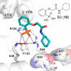
Binding of phosphonate inhibitor 19 (cyan sticks) to 14-3-3σ (white cartoon, sticks and surface). Residues from 14-3-3σ important for accommodation of 19 are shown as sticks; polar interactions are depicted as dotted black lines, and the semitransparent surface represents hydrophobic contacts (PDB ID: 4DHT).
Figure 13
Figure 13
Chemical structure of molecular tweezer 20 (A) and a 3D view of the molecule conformation it adopts for protein recognition (B). (C) Binding of molecular tweezer 20 (yellow sticks) to Lys214 of 14-3-3σ (white sticks) and the electron density (blue mesh, 2FO-FC, contoured at 1.0 σ). (D) Superimposition of the binding of molecular tweezer 20 (yellow spheres) and the ExoS peptide (416–430, purple sticks) to 14-3-3σ (white surface). (E) Molecular tweezer 20 (yellow sticks and surface) binding to Lys214 of 14-3-3σ (white cartoon and sticks) (PDB IDs: 4HQW and 4HRU).
Figure 14
Figure 14
Fusicoccane analogues, natural (21, and 22) or semi-synthetic (23, 24, and 25) that act as “molecular glue” model for stabilizing 14-3-3 binary structures.,,−
Figure 15
Figure 15
Semisynthetic derivatives 23, 26, and 27 stabilize the interaction between 14-3-3σ and TASK3 peptide. (A) Semisynthetic derivative 23 (purple sticks) and the C-terminus of TASK3 peptide (yellow sticks) in the binding groove of 14-3-3σ (cyan surface). (B) Electron density (red, blue, and black mesh, 2FO-FC, contoured at 1.0 σ) around 23 (purple sticks), C-terminus of TASK3 peptide (yellow sticks), and 14-3-3σ (green sticks). (C) Comparison of 23 (purple sticks) with 26 (orange sticks) and 27 (yellow sticks) in the binding pocket formed by 14-3-3σ (cyan surface) and TASK3 peptide (yellow surface) (PDB IDs: 3SMN, 3SMM, and 3SP5).
Figure 16
Figure 16
Comparison among 27, 26, and 23 in the stabilization of a “mode III” binder (TASK3) and “mode I/II” binder (C-Raf). (A) Overlay of 27 (green sticks), 26 (orange sticks), and 23 (purple stick) in the 14-3-3σ/TASK3 peptide (yellow sticks) complex. (B) The C-ring of 27 does not clash with “mode I/II” C-Raf peptide. (C) The hydroxylation of C12 in 26 clashes with the carbonyl oxygen of C-Raf P260. (D) The additional ring D of 23 clashes with both the carbonyl oxygen and the side group of C-Raf P260 (PDB IDs: 3SP5, 3SMM, 3SMN, and 4IEA).,
Figure 17
Figure 17
Compound 29 and derivative 30 stabilize the interaction between 14-3-3 and PMA2. (A) Chemical structures of 2830. (B) Compound 29 (yellow spheres) in the binding groove of T14-3-3e (green surface) having contact with PMA2 CT30 (blue surface). (C) Close-up of the T14-3e/PMA2/29 (green surface/blue surface/yellow sticks) interaction showing the electron density of 29 (gray mesh, 2FO-FC, contoured at 1.0 σ; PDB ID: 3M51).,
Figure 18
Figure 18
Crystal structure of the 14-3-3β/ChREBP/31 (AMP) complex. (A) Overview of ChREBP (purple cartoon) and 31 (yellow sticks and semitransparent spheres) bound to a monomer of 14-3-3β (solid white surface). (B) Detailed view of the contacts between 31 (yellow sticks), ChREBP (purple cartoon and sticks), and 14-3-3β (white cartoon and sticks). Polar contacts are depicted as black dotted lines (PDB ID: 5F74).

References

    1. Hatzivassiliou G.; Song K.; Yen I.; Brandhuber B. J.; Anderson D. J.; Alvarado R.; Ludlam M. J.; Stokoe D.; Gloor S. L.; Vigers G.; Morales T.; Aliagas I.; Liu B.; Sideris S.; Hoeflich K. P.; Jaiswal B. S.; Seshagiri S.; Koeppen H.; Belvin M.; Friedman L. S.; Malek S. RAF Inhibitors Prime Wild-Type RAF to Activate the MAPK Pathway and Enhance Growth. Nature 2010, 464 (7287), 431–435. 10.1038/nature08833. - DOI - PubMed
    1. Poulikakos P. I.; Persaud Y.; Janakiraman M.; Kong X.; Ng C.; Moriceau G.; Shi H.; Atefi M.; Titz B.; Gabay M. T.; Salton M.; Dahlman K. B.; Tadi M.; Wargo J. A.; Flaherty K. T.; Kelley M. C.; Misteli T.; Chapman P. B.; Sosman J. A.; Graeber T. G.; Ribas A.; Lo R. S.; Rosen N.; Solit D. B. RAF Inhibitor Resistance Is Mediated by Dimerization of Aberrantly Spliced BRAF(V600E). Nature 2011, 480 (7377), 387–390. 10.1038/nature10662. - DOI - PMC - PubMed
    1. Heidorn S. J.; Milagre C.; Whittaker S.; Nourry A.; Niculescu-Duvas I.; Dhomen N.; Hussain J.; Reis-Filho J. S.; Springer C. J.; Pritchard C.; Marais R. Kinase-Dead BRAF and Oncogenic RAS Cooperate to Drive Tumor Progression through CRAF. Cell 2010, 140 (2), 209–221. 10.1016/j.cell.2009.12.040. - DOI - PMC - PubMed
    1. Hopkins A. L.; Groom C. R. The Druggable Genome. Nat. Rev. Drug Discovery 2002, 1 (9), 727–730. 10.1038/nrd892. - DOI - PubMed
    1. Overington J. P.; Al-Lazikani B.; Hopkins A. L. How Many Drug Targets Are There?. Nat. Rev. Drug Discovery 2006, 5 (12), 993–996. 10.1038/nrd2199. - DOI - PubMed

Publication types

Substances